Semi-automatic Measurement of Ocular Volume from Facial Computed Tomography and Correlation with Axial Length
- Affiliations
-
- 1Department of Ophthalmology, Kyung Hee University Hospital at Gangdong, Kyung Hee University School of Medicine, Seoul, Korea. pbloadsky@naver.com
- 2Department of Ophthalmology, Kyung Hee University Medical Center, Kyung Hee University School of Medicine, Seoul, Korea.
- 3Department of Bioengineering, Kyung Hee University School of Medicine, Seoul, Korea.
- KMID: 2440447
- DOI: http://doi.org/10.3341/jkos.2019.60.3.210
Abstract
- PURPOSE
To measure the ocular volume from facial computed tomography (CT) scans using a semi-automatic computer program, and to analyze possible correlations between the axial length and ocular volume using regression analysis.
METHODS
Forty eyes from 20 facial CT scans were used to measure the ocular volumes. The cross-sectional ocular areas were calculated using a semi-automatic program based on MATLAB r2009a (MathWorks, Inc., Natick, MA, USA), and the ocular volumes were calculated from serial cross-sectional areas. The axial lengths were measured by A-scan ultrasound. Statistical analysis including regression analysis was used to determine possible correlations between the ocular volumes and axial lengths.
RESULTS
The mean ocular volumes measured in males and females were 7.16 ± 1.80 cm3 and 7.24 ± 3.38 cm3, respectively. The mean axial lengths measured in males and females were 23.47 ± 0.69 mm and 23.23 ± 1.64 mm, respectively. There were positive correlations using Pearson's correlation coefficient and the partial correlation coefficient adjusted by axial length. Using regression analysis, the following statistically significant equation was derived: (ocular volume [cm3] = 0.0056558 × axial length3 [mm3] − 0.1798106 × axial length2 [mm2] + 32.9008570 [p < 0.001, R2 = 0.384]).
CONCLUSIONS
The ocular volume measurement tool in this study was noninvasive and very useful, without special equipment. Accurate estimation of ocular volumes by a statistical equation was feasible, and these findings may be helpful in further study of various ocular diseases and in predicting preoperative and postoperative ocular volumes.
Figure
-
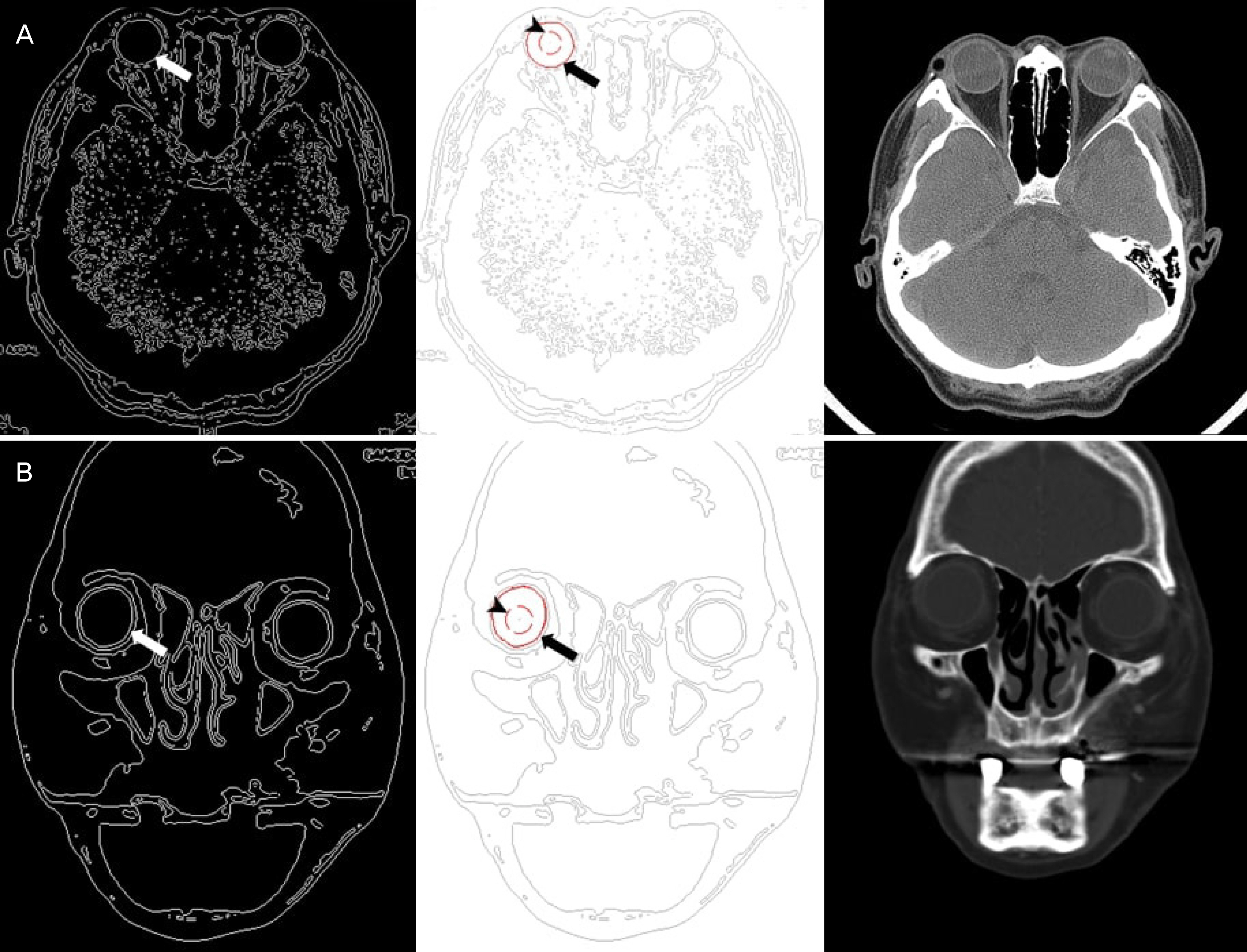
Figure 1. The process of measurement of cross-sectional areas from axial scans (A) and from coronal scans (B). The white arrows are ocular contours which were automatically displayed by the program. The seed points are marked with arrowheads and the results of automated measurement are marked with black arrows. This process was repeated in all scan sections that show an ocular contours. Each measured cross-sectional area was calculated as a volume through a conversion formula.

Figure 2. Flow chart of measuring ocular volume from facial computed tomography using MATLAB (MATLAB r2009a, MathWorks, Inc., Natick, MA, USA) based semi-automatic program. Otsu's algorithm and snake algorithm were used to detect eyeball and to check eyeball anatomy.
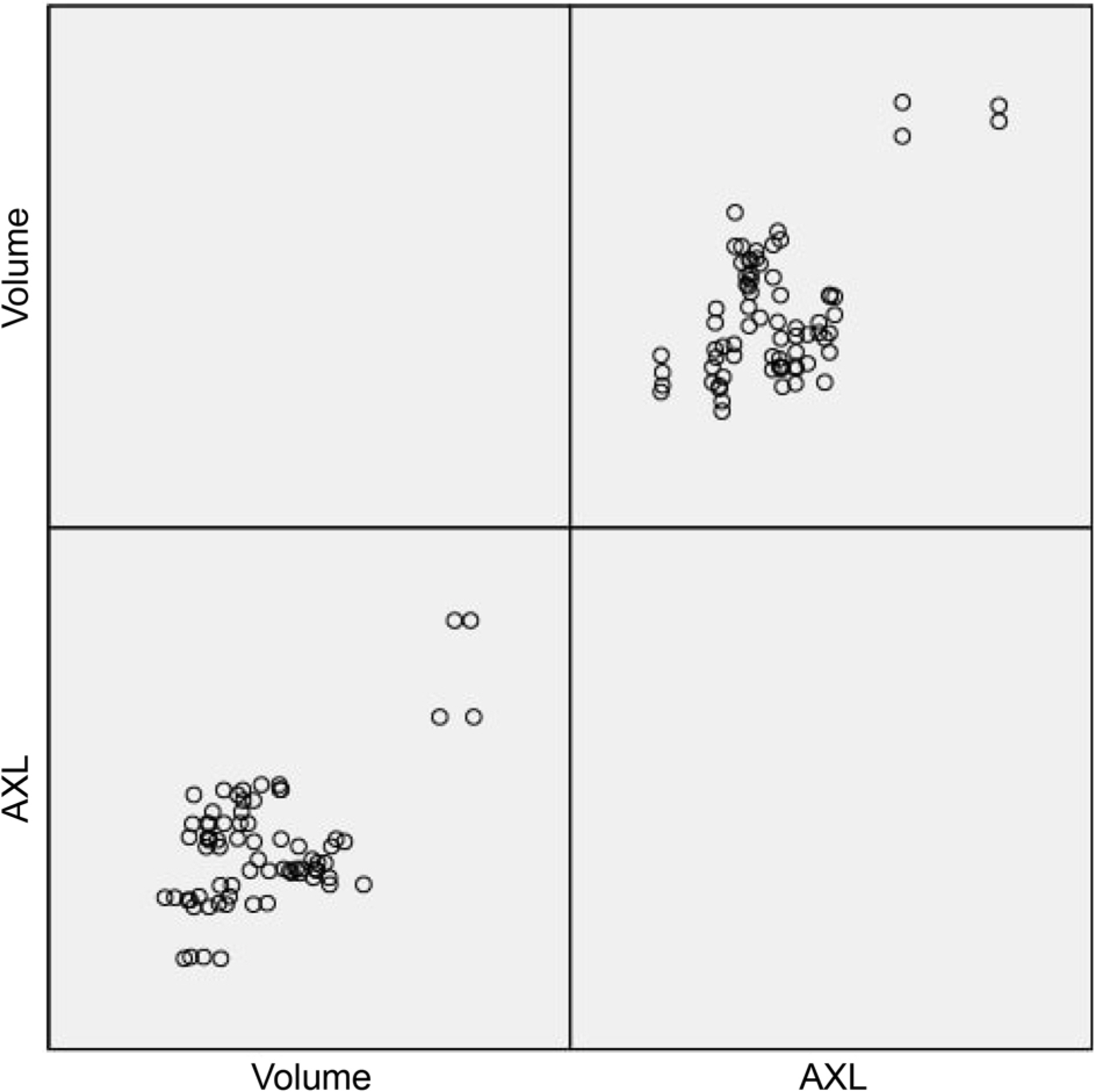
Figure 3. Scatter plot of ocular volume (volume, cm3) and AXL (mm). There were positive correlations in Pearson correlation coefficient and partial correlation coefficient adjusted by AXL. AXL = axial length.
Reference
-
References
1. Lim LS, Chong GH, Tan PT, et al. Distribution and determinants of eye size and shape in newborn children: a magnetic resonance imaging analysis. Invest Ophthalmol Vis Sci. 2013; 54:4791–7.
Article2. Liebovitch LS. Why the eye is round. Fischbarg J, editor. Advances in Organ Biology. Elsevier;2005. chap. 10.
Article3. Lee JH. Experimental scleral buckling reduction of ocular volume by scleral buckles. J Korean Ophthalmol Soc. 1980; 21:181–4.4. Park SY, Choi KS. Intraocular pressure elevation after intravitreal triamcinolone acetonide of different volumes: comparing 0.1 ml vs 0.05 ml. J Korean Ophthalmol Soc. 2008; 49:589–94.
Article5. Adelman RA, Zheng Q, Mayer HR. Persistent ocular hypertension following intravitreal bevacizumab and ranibizumab injections. J Ocul Pharmacol Ther. 2010; 26:105–10.
Article6. Cacciamani A, Oddone F, Parravano M, et al. Intravitreal injection of bevacizumab: changes in intraocular pressure related to ocular axial length. Jpn J Ophthalmol. 2013; 57:63–7.
Article7. Kotliar K, Maier M, Bauer S, et al. Effect of intravitreal injections and volume changes on intraocular pressure: clinical results and abdominal model. Acta Ophthalmol Scand. 2007; 85:777–81.8. Kim SW, Oh J, Yang KS, et al. Risk factors for the development of transient hypotony after silicone oil removal. Retina. 2010; 30:1228–36.
Article9. Ha SW, Kwon SJ, Park DH, Shin JP. Transient hypotony after abdominal oil removal in rhegmatogenous retinal detachment. J Korean Ophthalmol Soc. 2013; 54:85–91.10. Catalu CT, Istrate SL, Voinea LM, et al. Ocular implants-methods of ocular reconstruction following radical surgical interventions. Rom J Ophthalmol. 2018; 62:15–23.11. Wales RC. Ocular measurement by simple gravimetric methods. Invest Ophthalmol Vis Sci. 1977; 16:580–2.12. Igbinedion BO, Ogbeide OU. Measurement of normal ocular abdominal by the use of computed tomography. Niger J Clin Pract. 2013; 16:315–9.13. Nagra M, Gilmartin B, Logan NS. Estimation of ocular volume from axial length. Br J Ophthalmol. 2014; 98:1697–701.
Article14. Choi JH, Park IK, Choi SJ, Shin JH. Measurement of orbital abdominal from facial CT scans using a semi-automatic computer program. J Korean Ophthalmol Soc. 2015; 56:168–73.15. Tomlinson A, Phillips CI. Applanation tension and axial length of the eyeball. Br J Ophthalmol. 1970; 54:548–53.
Article16. Bekerman I, Gottlieb P, Vaiman M. Variations in eyeball diameters of the healthy adults. J Ophthalmol. 2014; 2014:503645.
Article17. Kim NS, Choi YJ, Hong YJ. The anterior chamber depth according to age in normal urban adult. J Korean Ophthalmol Soc. 1995; 36:1193–8.18. Huang Q, Huang Y, Luo Q, Fan W. Ocular biometric characteristics of cataract patients in western China. BMC Ophthalmology. 2018; 18:99.
Article19. Morris HJ, Tang J, Cruz Perez B, et al. Correlation between abdominal responses of posterior sclera and IOP elevations during micro intraocular volume change. Invest Ophthalmol Vis Sci. 2013; 54:7215–22.20. Bayoudh W, Carstesen D, Walter P, Weinberger AWA. Intraocular silicone implant to treat chronic ocular hypotony: an in vivo trial. Graefes Arch Clin Exp Ophthalmol. 2017; 255:1947–55.
Article21. Ikuno Y. Overview of the complications of high myopia. Retina. 2017; 37:2347–51.
Article22. Wong YL, Saw SM. Epidemiology of pathologic myopia in asia and worldwide. Asia Pac J Ophthalmol (Phila). 2016; 5:394–402.
Article
- Full Text Links
-
- Actions
-
Cited
- CITED
-
- Close
- Share
-
- Similar articles
-
- Measurement of Orbital Volume from Facial CT Scans Using a Semi-Automatic Computer Program
- Measurement of Orbital Volume from Different Slice Thickness Facial Computed Tomography Scans Using a Semi-automatic Program
- A Case of Excessive Axial Length Elongation Following Retinal Detachment Vitrectomy
- Interrelationship of the Refractory Error and the Ocular Axial Length and the Anterior Chamber Depth in the Myopic Eyes
- The Relationship between Axial Length, Refractive Power and Foveal Thickness Measured by OCT in Koreans
