J Korean Ophthalmol Soc.
2015 Feb;56(2):168-173. 10.3341/jkos.2015.56.2.168.
Measurement of Orbital Volume from Facial CT Scans Using a Semi-Automatic Computer Program
- Affiliations
-
- 1Department of Ophthalmology, Kyung Hee University Hospital at Gangdong, Kyung Hee University School of Medicine, Seoul, Korea. pbloadsky@naver.com
- 2Department of Ophthalmology, Kyung Hee University Medical Center, Kyung Hee University School of Medicine, Seoul, Korea.
- 3Department of Bioengineering, Kyung Hee University School of Medicine, Seoul, Korea.
- KMID: 2215998
- DOI: http://doi.org/10.3341/jkos.2015.56.2.168
Abstract
- PURPOSE
To measure the orbital volume from facial CT scans using a semi-automatic computer program.
METHODS
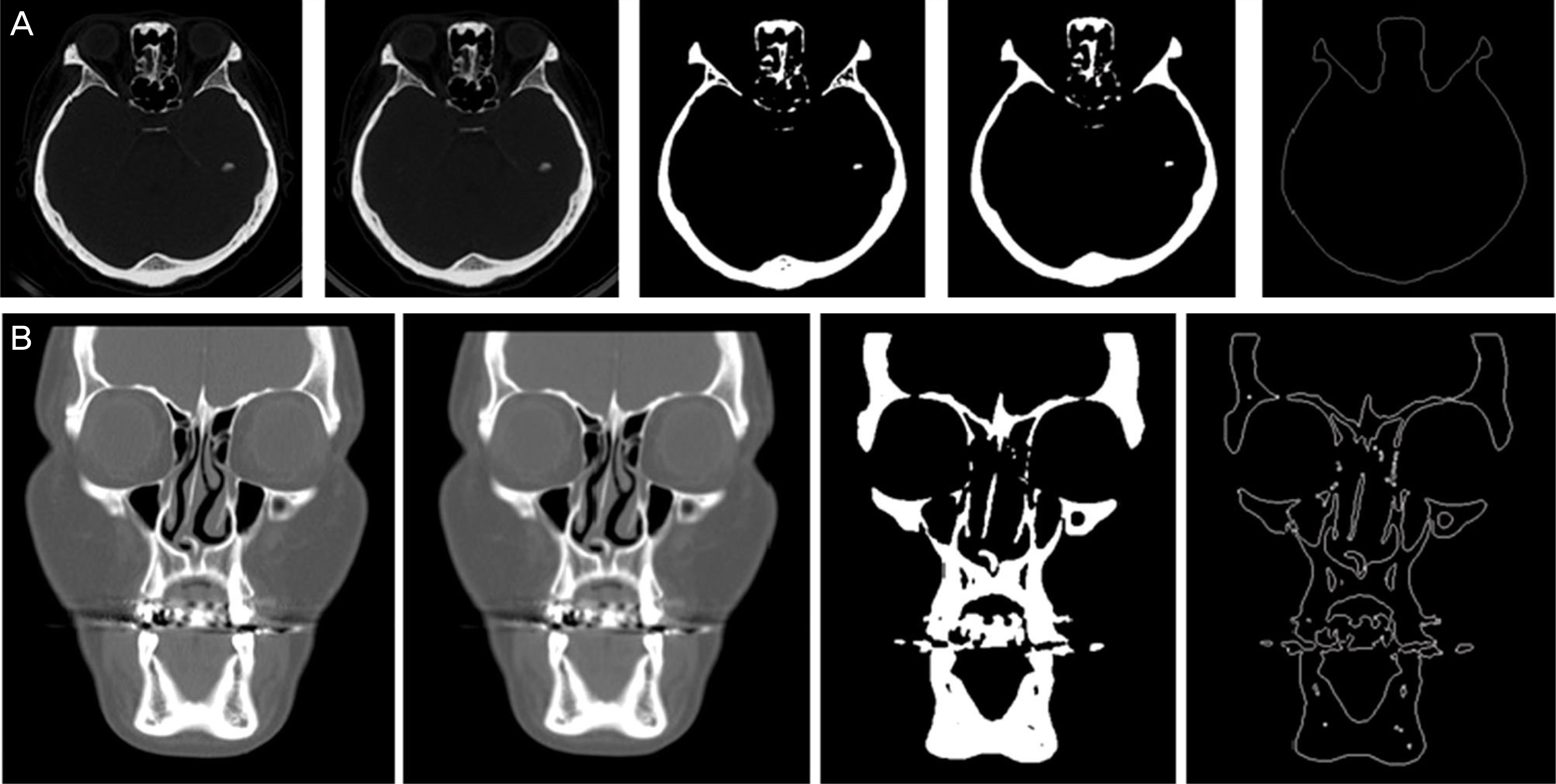
Axial and coronal slices of 35 facial CT scans were used to measure the orbital volume. The cross-sectional area was determined from each slice using a semi-automated computer program (MATLAB 2009a). Next, the orbital volume was calculated from serial reconstruction of the cross-sections.
RESULTS
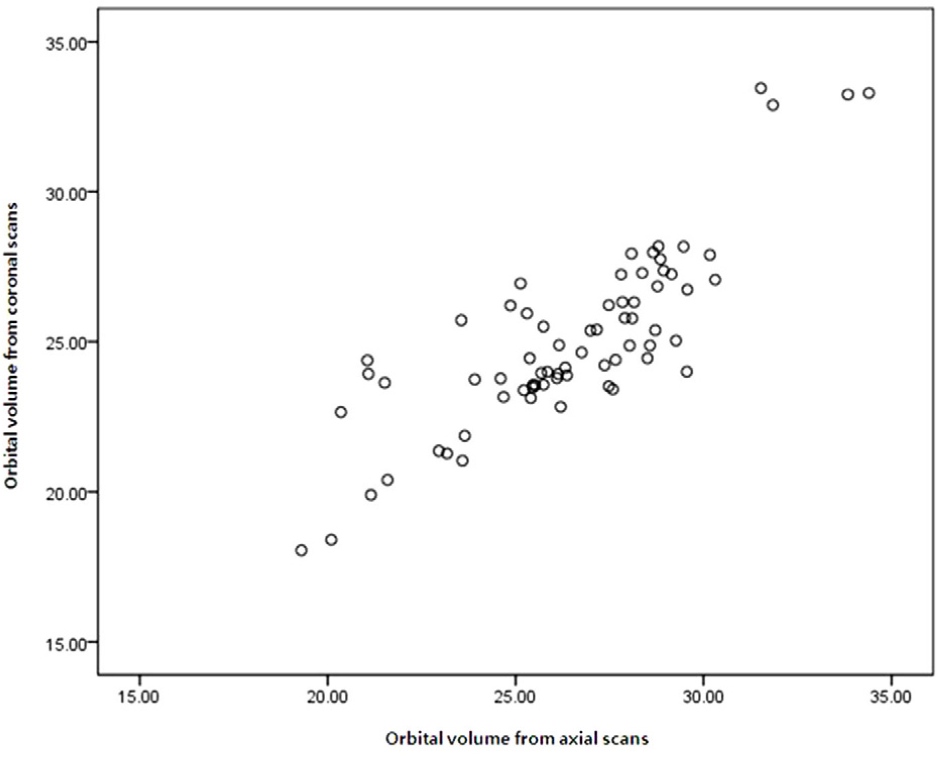
The measured value in males was 26.34 +/- 3.09 cm3 in the right orbit and 26.30 +/- 3.21 cm3 in the left orbit from axial scans, and 26.58 +/- 2.76 cm3 in the right orbit and 26.59 +/- 2.75 cm3 in the left orbit from coronal scans. In females, the values were 23.84 +/- 2.29 cm3 in the right orbit and 23.89 +/- 2.33 cm3 in the left orbit from axial scans, and 24.06 +/- 2.90 cm3 in the right orbit and 24.10 +/- 2.82 cm3 in the left orbit from coronal scans. There was high positive correlation (r = +0.832, p = 0.0001) in measured orbital volume between axial and coronal scans.
CONCLUSIONS
The orbital volume measurement from facial CT scans using a semi-automatic computer program is very useful. This method should prove useful in further studies examining the correlation of orbital volume variation in many ophthalmologic disorders.
Keyword
Figure
Reference
-
References
1. Phillips PH. The orbit. Ophthalmol Clin North Am. 2001; 14:109–27. viii.2. Cooper WC. A method for volume determination of the orbit and its contents by high resolution axial tomography and quantitative digital image analysis. Trans Am Ophthalmol Soc. 1985; 83:546–609.3. Forbes G, Gehring DG, Gorman CA. . Volume measurements of normal orbital structures by computed tomographic analysis. AJR Am J Roentgenol. 1985; 145:149–54.
Article4. Kwon J, Barrera JE, Most SP. Comparative computation of orbital volume from axial and coronal CT using three-dimensional image analysis. Ophthal Plast Reconstr Surg. 2010; 26:26–9.
Article5. Bite U, Jackson IT, Forbes GS, Gehring DG. Orbital volume measurements in enophthalmos using three-dimensional CT imaging. Plast Reconstr Surg. 1985; 75:502–8.
Article6. Kim TH, Jun HS, Byun YJ. The normal value of adult Korean orbital volume in three-dimensional computerized tomography. J Korean Ophthalmol Soc. 2001; 42:1011–5.7. Byun YG, Kim YI. High resolution satellite image segmentation algorithm development using seed-based region growing. Journal of the Korean Society of Surveying, Geodesy, Photogrammetry and Cartography. 2010; 28:421–30.8. Hojjatoleslami SA, Kittler J. Region growing: a new approach. IEEE Trans Image Process. 1998; 7:1079–84.
Article9. Leymarie F, Levine MD. Tracking deformable objects in the plane using an active contour model. Pattern Analysis and Machine Intelligence, IEEE Transactions on. 1993; 15:617–34.
Article10. Xu C, Prince JL. Snakes, shapes, and gradient vector flow. IEEE Trans Image Process. 1998; 7:359–69.11. Ramieri G, Spada MC, Bianchi SD, Berrone S. Dimensions and volumes of the orbit and orbital fat in posttraumatic enophthalmos. Dentomaxillofac Radiol. 2000; 29:302–11.
Article12. Furuta M. Measurement of orbital volume by computed tomography: especially on the growth of the orbit. Jpn J Ophthalmol. 2001; 45:600–6.
Article13. Forbes G, Gorman CA, Gehring D, Baker HL Jr. Computer analysis of orbital fat and muscle volumes in Graves ophthalmopathy. AJNR Am J Neuroradiol. 1983; 4:737–40.14. Chapter 32 Embryology and Anatomy of the Orbit and Lacrimal System. Tasman W, Jaeger EA, editors. Duane's Ophthalmology. Lippincott/Williams & Wilkins;2007.15. Acer N, Sahin B, Ergür H. . Stereological estimation of the orbital volume: a criterion standard study. J Craniofac Surg. 2009; 20:921–5.16. Trokel SL, Jakobiec FA. Correlation of CT scanning and pathologic features of ophthalmic Graves' disease. Ophthalmology. 1981; 88:553–64.
Article17. Whitehouse RW, Jackson A. Measurement of orbital volumes following trauma using lowdose computed tomography. Eur Radiol. 1993; 3:145–9.
Article
- Full Text Links
- Actions
-
Cited
- CITED
-
- Close
- Share
- Similar articles
-
- Measurement of Orbital Volume from Different Slice Thickness Facial Computed Tomography Scans Using a Semi-automatic Program
- Semi-automatic Measurement of Ocular Volume from Facial Computed Tomography and Correlation with Axial Length
- The Normal Value of Adult Korean Orbital Volume in Three-Dimensional Computerized Tomography
- Validity of Posterior Anterior Cephalometric and 3D-CT for Orbital Canting Analysis
- Quantitative Analysis of the Orbital Volume Change in Isolated Zygoma Fracture


