Ann Dermatol.
2019 Apr;31(2):209-212. 10.5021/ad.2019.31.2.209.
Symmetrical Giant Facial Plaque-Type Juvenile Xanthogranuloma: A Case Report with a Successful Response to Fractional COâ‚‚ Laser Treatment
- Affiliations
-
- 1Department of Dermatology, Severance Hospital, Cutaneous Biology Research Institute, Yonsei University College of Medicine, Seoul, Korea. ODDUNG93@yuhs.ac
- KMID: 2439068
- DOI: http://doi.org/10.5021/ad.2019.31.2.209
Abstract
- Symmetrical giant facial plaque-type juvenile xanthogranuloma (SGFP-JXG) is a rare variant of juvenile xanthogranuloma, reported only in two cases in the literature. We report a case of a 3-year-old Korean boy who developed bilateral yellowish indurated plaques on both cheeks since 1 year after birth. A skin biopsy revealed numerous foam cells and Touton type giant cells throughout the upper dermis, and its immunohistochemical studies resulted positive for CD68 and negative for S-100. The boy was therefore diagnosed as a persistent SGFP-JXG. As the lesion did not show any signs of spontaneous regression, we performed a single session of fractional ablative COâ‚‚ laser, which resulted in a significant reduction of the lesion. This is the first case report of a persistent SGFP-JXG on which a single ablative laser therapy was performed with a successful outcome.
MeSH Terms
Figure
-
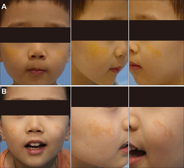
Fig. 1 We received the patient's consent form for publishing all photographic materials. (A) Initial photography of the bilateral symmetric yellowish plaques distributed on both cheeks. (B) Six-month follow-up photography after a single session of fractional ablative CO2 laser, showing reduced sizes of the lesions.
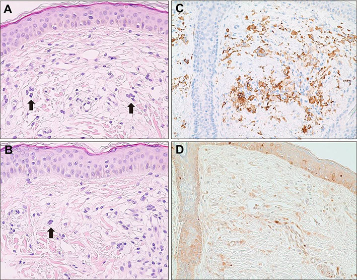
Fig. 2 (A, B) Haematoxylin-eosin staining of the yellowish plaque on the right cheek. Numerous foam cells and Touton giant cells with occasional foreign body giant cells are seen as indicated by arrows. (C) Immunohistochemical staining of the lesion showing CD68 positive histiocytes. (D) Immunohistochemical staining of the lesion showing a negative staining result on S-100 (×20).
Reference
-
1. Gunson TH, Birchall NM. Symmetrical giant facial plaque-type juvenile xanthogranuloma. J Am Acad Dermatol. 2008; 59:2 Suppl 1. S56–S57.
Article2. Szczerkowska-Dobosz A, Kozicka D, Purzycka-Bohdan D, Biernat W, Stawczyk M, Nowicki R. Juvenile xanthogranuloma: a rare benign histiocytic disorder. Postepy Dermatol Alergol. 2014; 31:197–200.3. Dehner LP. Juvenile xanthogranulomas in the first two decades of life: a clinicopathologic study of 174 cases with cutaneous and extracutaneous manifestations. Am J Surg Pathol. 2003; 27:579–593.
Article4. Sangüeza OP, Salmon JK, White CR Jr, Beckstead JH. Juvenile xanthogranuloma: a clinical, histopathologic and immunohistochemical study. J Cutan Pathol. 1995; 22:327–335.
Article5. Yazganoglu KD, Erdem Y, Buyukbabani N, Baykal C. A giant congenital plaque. Pediatr Dermatol. 2012; 29:217–218.
Article6. Sugiura K, Hasegawa Y, Shimoyama Y, Hashizume H, Akiyama M. Symmetrical giant facial plaque-type juvenile xanthogranuloma persisting beyond 10 years of age. Acta Derm Venereol. 2014; 94:465–466.
Article7. Miyagawa F, Fukumoto T, Kobayashi N, Asada H. Successful treatment of diffuse normolipemic plane xanthoma with probucol. Case Rep Dermatol. 2013; 5:148–151.
Article8. Altman J, Winkelmann RK. Diffuse normolipemic plane xanthoma. Generalized xanthelasma. Arch Dermatol. 1962; 85:633–640.9. Oka M, Okamura A, Kawano S, Fukumoto T, Sakaguchi M, Nishigori C. Diffuse plane normolipemic xanthoma associated with chronic myelomonocytic leukemia-1. Eur J Dermatol. 2014; 24:112–113.
Article10. Vail JT Jr, Adler KR, Rothenberg J. Cutaneous xanthomas associated with chronic myelomonocytic leukemia. Arch Dermatol. 1985; 121:1318–1320.
Article11. Kim KJ, Lee DP, Suh HS, Lee MW, Choi JH, Moon KC, et al. Diffuse plane xanthoma in a patient with chronic myeloid leukemia. J Dermatol. 2004; 31:503–505.
Article12. Lorenz S, Hohenleutner S, Hohenleutner U, Landthaler M. Treatment of diffuse plane xanthoma of the face with the Erbium:YAG laser. Arch Dermatol. 2001; 137:1413–1415.
Article13. Bragg J. Diffuse plane xanthomata. Dermatol Online J. 2005; 11:4.
Article14. Klemke CD, Held B, Dippel E, Goerdt S. Multiple juvenile xanthogranulomas successfully treated with CO laser. J Dtsch Dermatol Ges. 2007; 5:30–33.15. Trelles MA, Leclère FM, Martínez-Carpio PA. Fractional carbon dioxide laser and acoustic-pressure ultrasound for transepidermal delivery of cosmeceuticals: a novel method of facial rejuvenation. Aesthetic Plast Surg. 2013; 37:965–972.
Article
