Invasive Micropapillary Carcinoma in Axillary Ectopic Breast and Synchronous Ductal Carcinoma In Situ in the Contralateral Breast
- Affiliations
-
- 1Department of Radiology, Chonnam National University Hwasun Hospital, Chonnam National University Medical School, Hwasun, Korea. nico1220@dreamwiz.com
- 2Department of Pathology, Chonnam National University Hwasun Hospital, Chonnam National University Medical School, Hwasun, Korea.
- 3Department of Surgery, Chonnam National University Hwasun Hospital, Chonnam National University Medical School, Hwasun, Korea.
- KMID: 2439000
- DOI: http://doi.org/10.4048/jbc.2017.20.3.314
Abstract
- The development of ectopic breast tissue is attributable to the failure of primitive mammary tissue to regress after the development of the mammary ridge, except at pectoral breast sites, and is most often evident in the axillae. Several benign and malignant breast diseases have been reported in ectopic axillary breast tissues. The most common cancerous pathology of ectopic breast tissue is invasive ductal carcinoma. Ectopic breast cancer presenting with simultaneous primary cancer of the pectoral breast is extremely rare. Herein, we report an invasive micropapillary carcinoma of an axillary ectopic breast, combined with a synchronous ductal carcinoma in situ in the contralateral pectoral breast of a 61-year-old woman.
Keyword
MeSH Terms
Figure
-
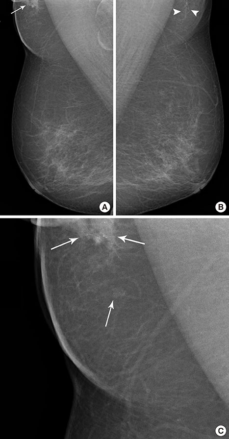
Figure 1 Mammographic findings of invasive micropapillary carcinoma in axillary ectopic breast. (A) Mediolateral oblique view of mammogram shows focal asymmetry with microcalcifications (arrow) in the right axilla. (B) Axillary fibroglandular tissue with the same radiological appearance as normal breast tissue (arrowheads) is also seen in the left axilla. (C) Zoomed mediolateral oblique view of right mammogram shows focal asymmetry and mass with microcalcifications (arrows) in the axilla.
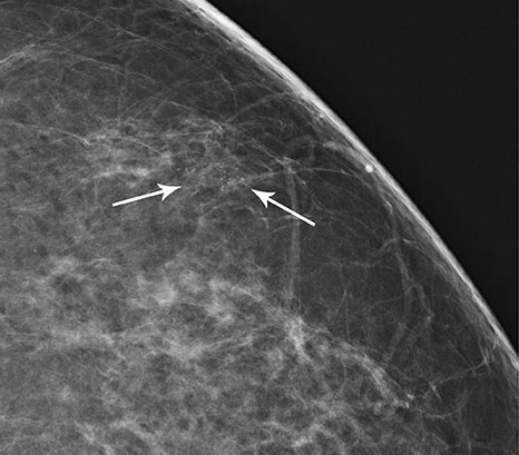
Figure 2 Mammographic findings of left breast. Magnified craniocaudal view of mammogram shows grouped, fine pleomorphic microcalcifications (arrows) in the upper outer quadrant of the left breast.
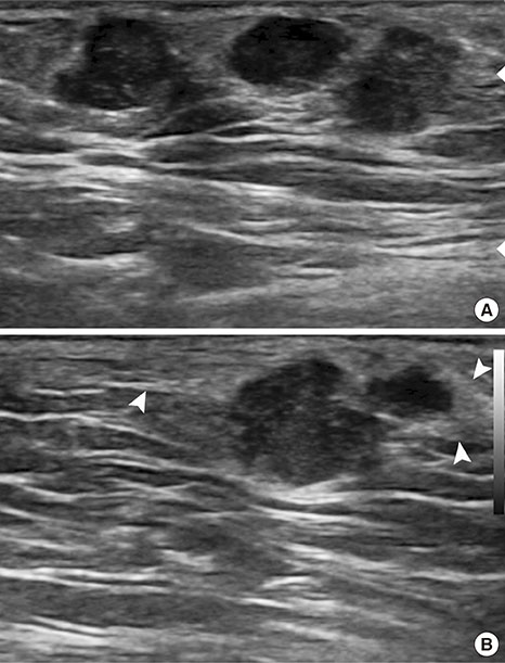
Figure 3 Sonographic findings of invasive micropapillary carcinoma in axillary ectopic breast. (A) Ultrasound image obtained in a local breast clinic shows several, irregular hypoechoic masses in the right axilla. (B) Echogenic area with the same appearance as that of normal breast tissue compatible with ectopic breast (arrowheads) is seen around the masses.
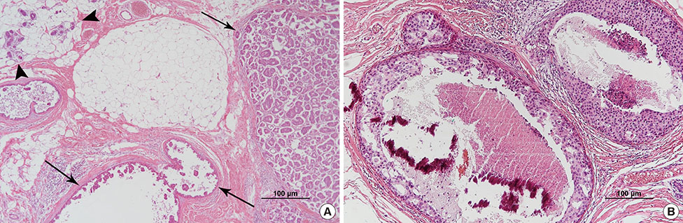
Figure 4 Histopathological findings. (A) Invasive micropapillary carcinoma and micropapillary ductal carcinoma in situ (arrows) are seen within the fatty mammary stroma with mammary lobules (arrowheads) in the right axilla (H&E stain, ×20). (B) Ductal carcinoma in situ with central necrosis and calcifications is seen in the left breast (H&E stain, ×200).
Reference
-
1. De Cholnoky T. Accessory breast tissue in the axilla. N Y State J Med. 1951; 51:2245–2248.2. Kim EY, Ko EY, Han BK, Shin JH, Hahn SY, Kang SS, et al. Sonography of axillary masses: what should be considered other than the lymph nodes? J Ultrasound Med. 2009; 28:923–939.3. Evans DM, Guyton DP. Carcinoma of the axillary breast. J Surg Oncol. 1995; 59:190–195.
Article4. Caceres M, Shih J, Eckert M, Gardner R. Metaplastic carcinoma in an ectopic breast. South Med J. 2002; 95:462–466.
Article5. Guerry RL, Pratt-Thomas HR. Carcinoma of supernumerary breast of vulva with bilateral mammary cancer. Cancer. 1976; 38:2570–2574.
Article6. Marshall MB, Moynihan JJ, Frost A, Evans SR. Ectopic breast cancer: case report and literature review. Surg Oncol. 1994; 3:295–304.
Article7. Hao JY, Yang CC, Liu FF, Yang YL, Li S, Li WD, et al. Accessory breast cancer occurring concurrently with bilateral primary invasive breast carcinomas: a report of two cases and literature review. Cancer Biol Med. 2012; 9:197–201.8. Alghamdi H, Abdelhadi M. Accessory breasts: when to excise? Breast J. 2005; 11:155–157.
Article9. Kitamura K, Kuwano H, Kiyomatsu K, Ikejiri K, Sugimachi K, Saku M. Mastopathy of the accessory breast in the bilateral axillary regions occurring concurrently with advanced breast cancer. Breast Cancer Res Treat. 1995; 35:221–224.
Article10. DeFilippis EM, Arleo EK. The ABCs of accessory breast tissue: basic information every radiologist should know. AJR Am J Roentgenol. 2014; 202:1157–1162.
Article11. Yerra L, Karnad AB, Votaw ML. Primary breast cancer in aberrant breast tissue in the axilla. South Med J. 1997; 90:661–662.
Article12. Nihon-Yanagi Y, Ueda T, Kameda N, Okazumi S. A case of ectopic breast cancer with a literature review. Surg Oncol. 2011; 20:35–42.
Article13. Kuroda H, Sakamoto G, Ohnisi K, Itoyama S. Clinical and pathologic features of invasive micropapillary carcinoma. Breast Cancer. 2004; 11:169–174.
Article14. Yun SU, Choi BB, Shu KS, Kim SM, Seo YD, Lee JS, et al. Imaging findings of invasive micropapillary carcinoma of the breast. J Breast Cancer. 2012; 15:57–64.
Article15. Copeland MM, Geschickter CF. Diagnosis and treatment of premalignant lesions of the breast. Surg Clin North Am. 1950; 30:1717–1741.
Article
- Full Text Links
-
- Actions
-
Cited
- CITED
-
- Close
- Share
-
- Similar articles
-
- Invasive Micropapillary Carcinoma of the Breast: A clinicopathologic study of 16 cases
- Invasive Lobular Carcinoma of the Breast Associated with Mixed Lobular and Ductal Carcinoma In Situ: A Case Report
- A Case Report on Ductal Carcinoma in situ Arising from Axillary Accessory Breast Tissue
- Concurrent Invasive Carcinoma and Fibroadenoma Arising from Bilateral Ectopic Breast Tissue in the Chest Wall: A Case Report and Literature Review
- Myoepithelial carcinoma with contralateral invasive micropapillary carcinoma of the breast
