Arthroscopic Repair of Acetabular Labral Tears Associated with Femoroacetabular Impingement: 7–10 Years of Long-Term Follow-up Results
- Affiliations
-
- 1Department of Orthopaedic Surgery, Chungnam National University School of Medicine, Daejeon, Korea. dshwang@cnu.ac.kr
- KMID: 2438331
- DOI: http://doi.org/10.4055/cios.2019.11.1.28
Abstract
- BACKGROUND
The purpose of this study is to report the long-term follow-up results of arthroscopic repair of acetabular labral tears with femoroacetabular impingement (FAI).
METHODS
Of 45 patients who underwent arthroscopic labral repair under the diagnosis of acetabular labral tears with FAI from January 2008 to December 2010 and met our inclusion criteria, 41 patients who were available for a long-term follow-up were included in the analysis. We compared the long-term follow-up results with the previously reported short-term follow-up results of the same patients. The mean follow-up period was 92.4 months (range, 85 to 117 months). There were 21 males and 20 females, and their mean age at surgery was 34.6 years (range, 16 to 54 years). A modified Harris hip score (mHHS), visual analog scale (VAS), hip outcome score-activity of daily living (HOS-ADL), hip outcome score-activity-sport-specific subscale (HOS-SSS), and patient satisfaction were used for evaluation of the clinical results and Tönnis grade for detection of early osteoarthritis (OA).
RESULTS
The mean VAS score decreased from 6.4 points to 1.8 points (p < 0.001), the mean mHHS increased from 59.5 points to 86.8 points (p < 0.001), and the mean HOS-ADL and HOS-SSS increased from 58.3 and 51.2, respectively, to 85.2 and 82.4, respectively (p < 0.001), between the preoperative and last follow-up assessment. The mean patient satisfaction score was 7.6 of 10. The average Tönnis grade at the last follow-up (0.67; range, 0 to 3) was not significantly different from the preoperative average (0.51; range, 0 to 1). Only one case was converted to total hip arthroplasty because of progression of OA at 8 years after surgery. Five cases of secondary arthroscopic surgery were performed before maximum 5 years postoperatively because of labro-synovial adhesion (three cases), pullout of the suture anchor (one case) or symptomatic heterotrophic ossification (one case).
CONCLUSIONS
The clinical and radiological long-term follow-up revealed that improvement after arthroscopic labral repair and osteoplasty for FAI were maintained in most cases without significant progression of arthritis. Anatomical recovery of the acetabular labrum was associated with the improvement of clinical symptoms.
MeSH Terms
Figure
-

Fig. 1 Visual analog scale (VAS) score and patient satisfaction score (A) and patient-reported outcome (B) collected preoperatively, at the short-term follow-up (FU; 2–5 years), and at the long-term FU (7–10 years). (C) Average Tönnis grade measured preoperatively and at the last long-term FU. (D) The graph shows changes in scores by the period (1: preoperative, 2: short-term FU, 3: long-term FU). mHHS: modified Harris hip score, HOS-ADL: hip outcome score-activity of daily living, HOS-SSS: hip outcome score-sport-specific subscale. *Scheffe's post hoc analysis, p < 0.005.

Fig. 2 A 47-year-old female had a right hip femoroacetabular impingement and acetabular labral tear. She underwent arthroscopic labral repair using two anchors and arthroscopic bumpectomy. (A) In arthroscopy, early degenerative change was shown in the femoral head cartilage (asterisk) and labral surface (arrow). (B) Acetabular cartilage surface fibrillation (asterisk) was observed. (C) She had a mild dysplastic hip on the right side and a Tönnis grade 1 osteoarthritis (arrow) in the preoperative period. (D) Joint space narrowing and degenerative changes developed gradually and Tönnis grade 3 (arrow) osteoarthritis was found at the last 86-month follow-up. (E) She received total hip arthroplasty.

Fig. 3 A 28-year-old female received arthroscopic labral repair and femoroplasty. After 2 months, she received revisional hip arthroscopy for pain and limited motions. (A) In arthroscopy, severe adhesion between the labrum (asterisk) and hip joint synovial capsule (arrow) was observed. (B) The simple frog-leg radiograph after primary arthroscopy showed incomplete femoroplasty on the femoral neck area (arrow) where the alpha angle was 64.5° and offset was 7.8 mm. (C) On the final frog-leg simple radiograph, we could confirm complete femoroplasty was performed: the alpha angle was 49.5° and offset was 10.2 mm (arrow) after revisional arthroscopy.
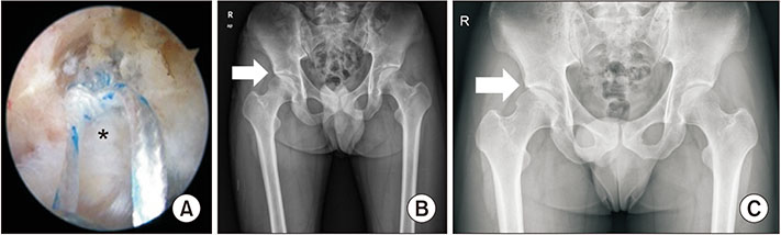
Fig. 4 (A) A 21-year-old male received arthroscopic labral repair (asterisk) and bumpectomy because of femoroacetabular impingement and labral tear in the right hip joint. (B) Preoperative simple radiogrph showing Tönnis grad 0 osteoarthritis (arrow) in the preoperative period. (C) Eight years later, the joint space became wider as he grew and the Tönnis grade was not changed (arrow).
Cited by 1 articles
-
Midterm-clinical Outcomes after Hip Arthroscopy in Middle-aged Patients with Early Osteoarthritis
Jeong-Kil Lee, Deuk-Soo Hwang, Chan Kang, Jung-Mo Hwang, Gi-Soo Lee, Long Zeng, Young-Cheol Park
Hip Pelvis. 2020;32(1):17-25. doi: 10.5371/hp.2020.32.1.17.
Reference
-
1. Takechi H, Nagashima H, Ito S. Intra-articular pressure of the hip joint outside and inside the limbus. Nihon Seikeigeka Gakkai Zasshi. 1982; 56(6):529–536.2. Kim YT, Azuma H. The nerve endings of the acetabular labrum. Clin Orthop Relat Res. 1995; (320):176–181.
Article3. Altenberg AR. Acetabular labrum tears: a cause of hip pain and degenerative arthritis. South Med J. 1977; 70(2):174–175.4. Czerny C, Hofmann S, Neuhold A, et al. Lesions of the acetabular labrum: accuracy of MR imaging and MR arthrography in detection and staging. Radiology. 1996; 200(1):225–230.
Article5. McCarthy JC, Noble PC, Schuck MR, Wright J, Lee J. The Otto E. Aufranc Award: the role of labral lesions to development of early degenerative hip disease. Clin Orthop Relat Res. 2001; (393):25–37.6. Ferguson SJ, Bryant JT, Ganz R, Ito K. The influence of the acetabular labrum on hip joint cartilage consolidation: a poroelastic finite element model. J Biomech. 2000; 33(8):953–960.
Article7. Ferguson SJ, Bryant JT, Ganz R, Ito K. The acetabular labrum seal: a poroelastic finite element model. Clin Biomech (Bristol, Avon). 2000; 15(6):463–468.
Article8. Ferguson SJ, Bryant JT, Ganz R, Ito K. An in vitro investigation of the acetabular labral seal in hip joint mechanics. J Biomech. 2003; 36(2):171–178.
Article9. Petersen W, Petersen F, Tillmann B. Structure and vascularization of the acetabular labrum with regard to the pathogenesis and healing of labral lesions. Arch Orthop Trauma Surg. 2003; 123(6):283–288.
Article10. Espinosa N, Rothenfluh DA, Beck M, Ganz R, Leunig M. Treatment of femoro-acetabular impingement: preliminary results of labral refixation. J Bone Joint Surg Am. 2006; 88(5):925–935.
Article11. Larson CM, Giveans MR, Stone RM. Arthroscopic debridement versus refixation of the acetabular labrum associated with femoroacetabular impingement: mean 3.5-year follow-up. Am J Sports Med. 2012; 40(5):1015–1021.
Article12. Philippon MJ, Ejnisman L, Ellis HB, Briggs KK. Outcomes 2 to 5 years following hip arthroscopy for femoroacetabular impingement in the patient aged 11 to 16 years. Arthroscopy. 2012; 28(9):1255–1261.
Article13. Klaue K, Durnin CW, Ganz R. The acetabular rim syndrome: a clinical presentation of dysplasia of the hip. J Bone Joint Surg Br. 1991; 73(3):423–429.
Article14. Suenaga E, Noguchi Y, Jingushi S, et al. Relationship between the maximum flexion-internal rotation test and the torn acetabular labrum of a dysplastic hip. J Orthop Sci. 2002; 7(1):26–32.
Article15. Lage LA, Patel JV, Villar RN. The acetabular labral tear: an arthroscopic classification. Arthroscopy. 1996; 12(3):269–272.
Article16. Kelly BT, Weiland DE, Schenker ML, Philippon MJ. Arthroscopic labral repair in the hip: surgical technique and review of the literature. Arthroscopy. 2005; 21(12):1496–1504.
Article17. Jackson TJ, Hanypsiak B, Stake CE, Lindner D, El Bitar YF, Domb BG. Arthroscopic labral base repair in the hip: clinical results of a described technique. Arthroscopy. 2014; 30(2):208–213.
Article18. Philippon MJ, Briggs KK, Yen YM, Kuppersmith DA. Outcomes following hip arthroscopy for femoroacetabular impingement with associated chondrolabral dysfunction: minimum two-year follow-up. J Bone Joint Surg Br. 2009; 91(1):16–23.19. Krych AJ, Thompson M, Knutson Z, Scoon J, Coleman SH. Arthroscopic labral repair versus selective labral debridement in female patients with femoroacetabular impingement: a prospective randomized study. Arthroscopy. 2013; 29(1):46–53.
Article20. Ayeni OR, Adamich J, Farrokhyar F, et al. Surgical management of labral tears during femoroacetabular impingement surgery: a systematic review. Knee Surg Sports Traumatol Arthrosc. 2014; 22(4):756–762.
Article21. Song Y, Ito H, Kourtis L, Safran MR, Carter DR, Giori NJ. Articular cartilage friction increases in hip joints after the removal of acetabular labrum. J Biomech. 2012; 45(3):524–530.
Article22. Jeon YS, Hwang DS, Kang C, Hwang JM, Lee GS. Arthroscopic labral repair associated with femoroacetabular impingement: short term 2-5 years follow-up results. Hip Pelvis. 2013; 25(2):115–120.
Article23. Kang C, Hwang DS, Jeon YS, Han SC, Lee GS, Kang DH. Arthroscopic treatment of femoroacetabular impingement of the hip: 5-7 years result. Hip Pelvis. 2012; 24(3):237–244.
Article24. Menge TJ, Briggs KK, Dornan GJ, McNamara SC, Philippon MJ. Survivorship and outcomes 10 years following hip arthroscopy for femoroacetabular impingement: labral debridement compared with labral repair. J Bone Joint Surg Am. 2017; 99(12):997–1004.
Article25. Hwang DS, Kang C, Cha SM, Kim JH. Second-look hip arthroscopy after arthroscopic labrectomy of the hip: preliminary report. J Korean Orthop Assoc. 2009; 44(4):480–485.
Article26. Gupta A, Redmond JM, Stake CE, Dunne KF, Domb BG. Does primary hip arthroscopy result in improved clinical outcomes?: 2-year clinical follow-up on a mixed group of 738 consecutive primary hip arthroscopies performed at a high-volume referral center. Am J Sports Med. 2016; 44(1):74–82.
Article27. Sawyer GA, Briggs KK, Dornan GJ, Ommen ND, Philippon MJ. Clinical outcomes after arthroscopic hip labral repair using looped versus pierced suture techniques. Am J Sports Med. 2015; 43(7):1683–1688.
Article28. Harris JD, McCormick FM, Abrams GD, et al. Complications and reoperations during and after hip arthroscopy: a systematic review of 92 studies and more than 6,000 patients. Arthroscopy. 2013; 29(3):589–595.
Article
- Full Text Links
-
- Actions
-
Cited
- CITED
-
- Close
- Share
-
- Similar articles
-
- Arthroscopic Labral Repair Associated with Femoroacetabular Impingement: Short Term 2-5 Years Follow-up Results
- Acetabular Labral Tears in Patients with Sports Injury
- Patient Satisfaction after Arthroscopic Repair of Acetabular Labral Tears
- Second-look Hip Arthroscopy after Arthroscopic Labrectomy of the Hip: Preliminary Report
- Ultrasonographic Usefulness for Diagnosis of Acetabular Labral Tear
