Ann Dermatol.
2012 May;24(2):228-229.
The Diagnostic Significance of Infiltration Pattern and Perilesional Lymphoid Cell Infiltrate in Dermatofibroma
- Affiliations
-
- 1Department of Dermatology, Kosin University College of Medicine, Busan, Korea. ksderm98@unitel.co.kr
Abstract
- No abstract available.
MeSH Terms
Figure
-
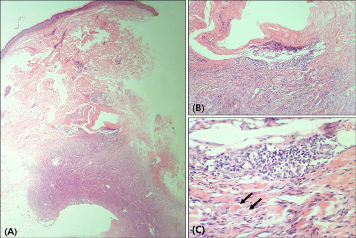
Fig. 1 (A) A well-circumscribed nodule deeply localized into the subcutaneous tissue (H&E, ×40). (B, C) Scattered collections of lymphocytes and sclerotic collagen (arrows) at the periphery of the tumor (H&E, B: ×100, C: ×400).
Reference
-
1. Han TY, Chang HS, Lee JH, Lee WM, Son SJ. A clinical and histopathological study of 122 cases of dermatofibroma (benign fibrous histiocytoma). Ann Dermatol. 2011. 23:185–192.
Article2. Kamino H, Jacobson M. Dermatofibroma extending into the subcutaneous tissue. Differential diagnosis from dermatofibrosarcoma protuberans. Am J Surg Pathol. 1990. 14:1156–1164.3. Gleason BC, Fletcher CD. Deep "benign" fibrous histiocytoma: clinicopathologic analysis of 69 cases of a rare tumor indicating occasional metastatic potential. Am J Surg Pathol. 2008. 32:354–362.
Article4. Zelger B, Sidoroff A, Stanzl U, Fritsch PO, Ofner D, Zelger B, et al. Deep penetrating dermatofibroma versus dermatofibrosarcoma protuberans. A clinicopathologic comparison. Am J Surg Pathol. 1994. 18:677–686.5. Harris RB, Duncan SC, Ecker RI, Winkelmann RK. Lymphoid follicles in subcutaneous inflammatory disease. Arch Dermatol. 1979. 115:442–443.
Article6. de Almeida LS, Requena L, Rütten A, Kutzner H, Garbe C, Pestana D, et al. Desmoplastic malignant melanoma: a clinicopathologic analysis of 113 cases. Am J Dermatopathol. 2008. 30:207–215.
Article
- Full Text Links
-
- Actions
-
Cited
- CITED
-
- Close
- Share
-
- Similar articles
-
- Two Patterns of Gastric Carcinoma with Lymphoid Stroma
- A Case of Dermatofibroma associated with Follicular Basal Cell Hyperplasia
- A Case of Primary Cutaneous Anaplastic Large Cell Lymphoma with Extensive Inflammatory Cell Infiltration
- Ki - 67 Expression in Cutaneous Lymphoid Infiltrates
- Two Cases of Dermatofibroma with Atrophic Features
