Magnified single-balloon enteroscopy in the diagnosis of intestinal follicular lymphoma: a case series
- Affiliations
-
- 1Department of Medicine, Shiga University of Medical Science, Otsu, Japan.
- 2Division of Clinical Nutrition, Shiga University of Medical Science, Otsu, Japan. sb@belle.shiga-med.ac.jp
- 3Department of Comprehensive Internal Medicine, Shiga University of Medical Science, Otsu, Japan.
- 4Division of Diagnostic Pathology, Shiga University of Medical Science, Otsu, Japan.
- 5Division of Digestive Endoscopy, Shiga University of Medical Science, Otsu, Japan.
- KMID: 2434165
- DOI: http://doi.org/10.5217/ir.2018.00003
Abstract
- The objective of this study was to evaluate the magnified endoscopic findings in the diagnosis of follicular lymphoma in the small intestine in comparison with those of intestinal follicular lymphoma and lymphangiectasia. Four patients with follicular lymphoma and 3 with lymphangiectasia in the small intestine were retrospectively analyzed. A prototype magnifying singleballoon enteroscope was used. The findings of the intestinal follicular lymphoma and lymphangiectasia were retrospectively analyzed to determine the magnified endoscopic findings of follicular lymphoma in the small intestine. Opaque white granules were observed in 3 of the 4 patients with follicular lymphoma. Magnified narrow-band imaging (NBI) of the opaque white granules showed stretched microvessels, which had a diminutive tree-like appearance. The remaining patient had no opaque white granules and only displayed whitish villi. Magnified NBI observation of the whitish villi revealed the absence of marginal villus epithelium, which was confirmed by histology. The magnified NBI enteroscopy revealed the diminutive tree-like appearance on the opaque white granules and the absence of marginal villus epithelium of the whitish villi in intestinal follicular lymphoma. These findings may be useful in diagnosing follicular lymphoma.
MeSH Terms
Figure
-
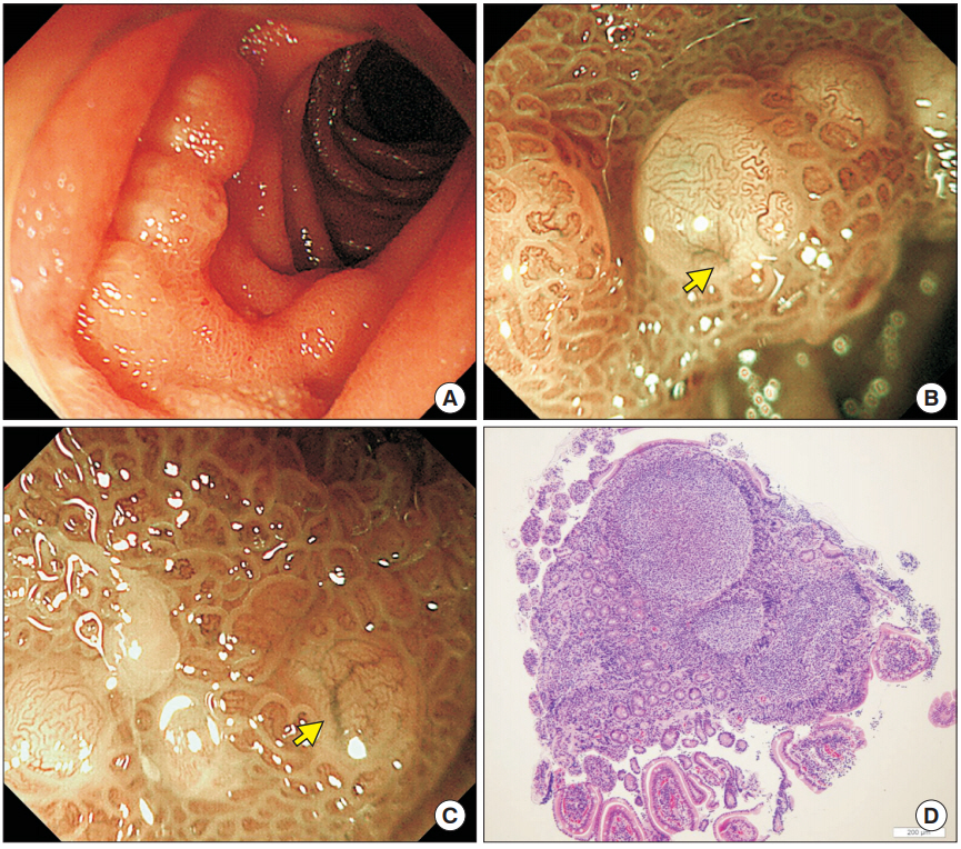
Fig. 1. Case 1: follicular lymphoma of the jejunum. (A) White light observation of the jejunal follicular lymphoma. (B, C) Narrowband imaging with magnified endoscopy revealed confluent hemispherical elevation without marginal villus epithelium. Diminutive tree-like appearance of microvessels (yellow arrow) and gyrus-like microvessels were observed. (D) Histological images of biopsy samples taken from the opaque white granules. Lymphoid follicles were present in the lamina propria and caused stretching of the epithelium (×40).
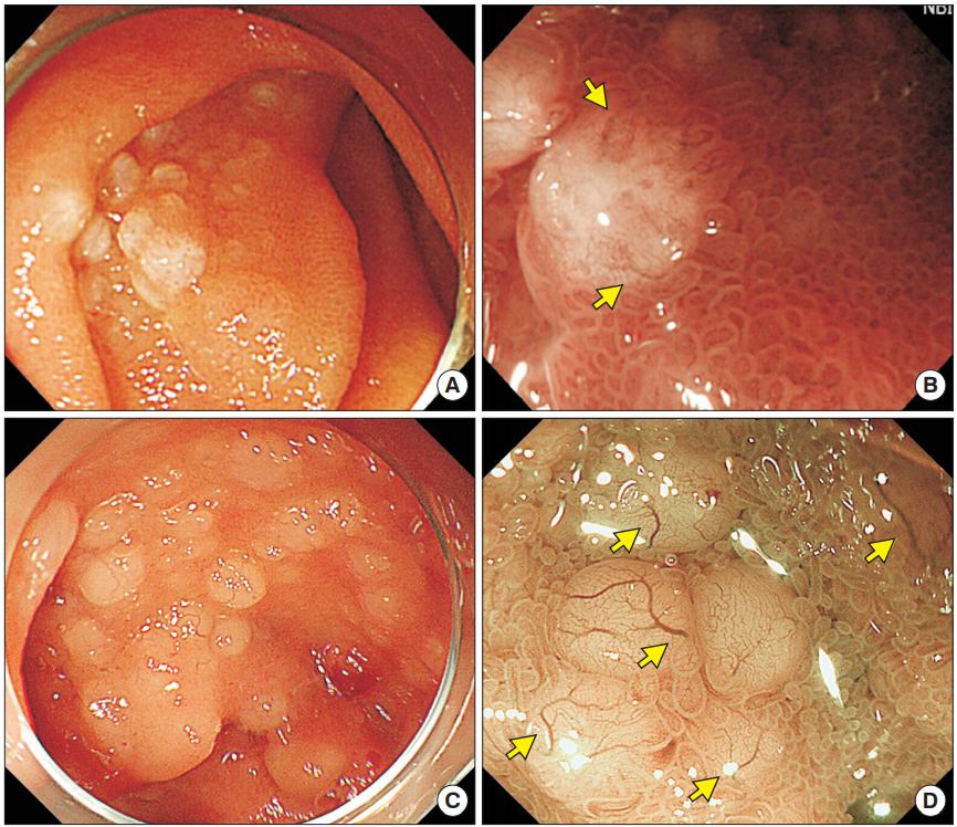
Fig. 2. Case 2 (A, B) and case 3 (C, D): follicular lymphoma of the jejunum. (A) White light observation of the jejunal follicular lymphoma. (B) Narrow-band imaging magnified endoscopy revealed confluent hemispherical elevation. Diminutive tree-like appearance (yellow arrows) was observed. (C) White light image of the jejunal follicular lymphoma. (D) Narrow-band imaging magnified observation revealed diminutive tree-like appearance (yellow arrows) on the opaque white granules.
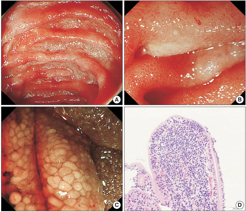
Fig. 3. Case 4: follicular lymphoma of the jejunum. (A) Jejunal lesion of follicular lymphoma seen on white light observation. (B) Magnified observation under white light demonstrated relatively homogenous whitish villi. (C) Narrow-band imaging magnified endoscopy demonstrated whitish villi without marginal villus epithelium. (D) Histological images of biopsy samples taken from whitish villi without marginal villus epithelium. Lymphoma cells infiltrated the lamina propria, and the epithelial cells were replaced by lymphoma cells at the tip of villi (×200).
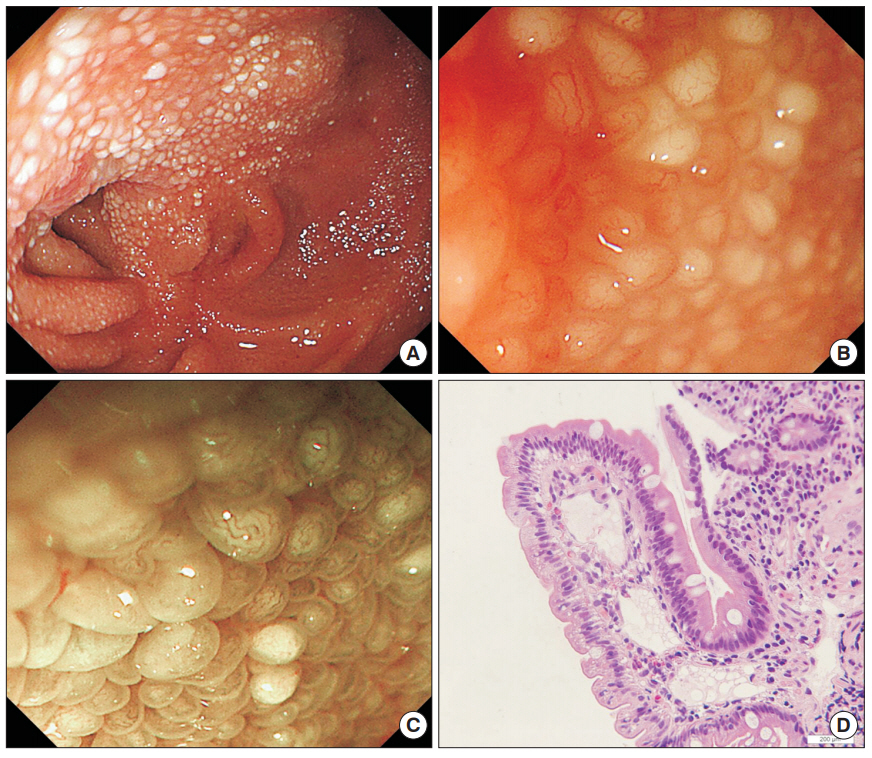
Fig. 4. Case 5: lymphangiectasia. (A) A patient with CD underwent single-balloon enteroscopy for endoscopic balloon dilatation for intestinal stricture. White light view of the ileum demonstrated a scar lesion and whitish villi in the ileum. (B, C) White light and narrow-band imaging magnified endoscopy revealed whitish villi with marginal villus epithelium. (D) Histological images of biopsy samples taken from the whitish villi. Markedly dilated lymphatics were present in the lamina propria (×200).
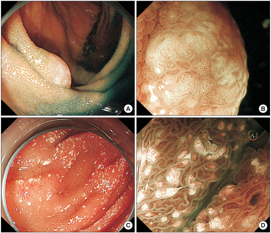
Fig. 5. Case 6 (A, B) and case 7 (C, D): lymphangiectasia. (A) The patient underwent single-balloon enteroscopy to identify the source of gastrointestinal bleeding. Lymphangioma was observed as a smooth elevated lesion with whitish villi. (B) Narrow-band imaging magnified endoscopy revealed enlarged whitish villi with gyruslike microvessels. (C) A patient who had undergone the Fontan procedure for single ventricle of right ventricular morphology, pulmonary artery occlusion and patent ductus arteriosus developed protein-losing enteropathy, and single-balloon enteroscopy was therefore performed. Scattered white spots were observed in the jejunum. (D) Narrow-band imaging magnified observation revealed scattered white spots within the tips of villi.
Reference
-
1. Yamamoto H, Sekine Y, Sato Y, et al. Total enteroscopy with a nonsurgical steerable double-balloon method. Gastrointest Endosc. 2001; 53:216–220.
Article2. Tsujikawa T, Saitoh Y, Andoh A, et al. Novel single-balloon enteroscopy for diagnosis and treatment of the small intestine: preliminary experiences. Endoscopy. 2008; 40:11–15.
Article3. Yamamoto S, Nakase H, Yamashita K, et al. Gastrointestinal follicular lymphoma: review of the literature. J Gastroenterol. 2010; 45:370–388.
Article4. Chowdhury M, Endo M, Chiba T, et al. Characterization of follicular lymphoma in the small intestine using double-balloon endoscopy. Gastroenterol Res Pract. 2009; 2009:835258.
Article5. Iwamuro M, Okada H, Kawano S, et al. A multicenter survey of enteroscopy for the diagnosis of intestinal follicular lymphoma. Oncol Lett. 2015; 10:131–136.
Article6. Inoue N, Isomoto H, Shikuwa S, Mizuta Y, Hayashi T, Kohno S. Magnifying endoscopic observation of primary follicular lymphoma of the duodenum by using the narrow-band imaging system. Gastrointest Endosc. 2009; 69:158–159.
Article7. Norimura D, Isomoto H, Imaizumi Y, et al. Case series of duodenal follicular lymphoma, observed by magnified endoscopy with narrow-band imaging. Gastrointest Endosc. 2011; 74:428–434.
Article8. Iwamuro M, Okada H, Takata K, et al. Magnified endoscopic features of duodenal follicular lymphoma and other whitish lesions. Acta Med Okayama. 2015; 69:37–44.9. Tada M, Suyama Y, Shimizu T, et al. Observation of the villi with the magnifying entero-colonoscopes. Gastroenterol Endosc. 1980; 22:647–654.10. Murata M, Bamba S, Takahashi K, et al. Application of novel magnified single balloon enteroscopy for a patient with Cronkhite-Canada syndrome. World J Gastroenterol. 2017; 23:4121–4126.
Article11. Asakura H, Miura S, Morishita T, et al. Endoscopic and histopathological study on primary and secondary intestinal lymphangiectasia. Dig Dis Sci. 1981; 26:312–320.
Article12. Iwamuro M, Okada H, Takata K, et al. Magnifying endoscopic observation of duodenal involvement of follicular lymphoma before and after chemotherapy. Intern Med. 2015; 54:1741–1745.13. Nonaka K, Ishikawa K, Arai S, et al. A case of gastric mucosaassociated lymphoid tissue lymphoma in which magnified endoscopy with narrow band imaging was useful in the diagnosis. World J Gastrointest Endosc. 2012; 4:151–156.
Article14. Iwamuro M, Okuda M, Yumoto E, et al. Magnifying endoscopy for intestinal follicular lymphoma is helpful for prompt diagnosis. Gut Liver. 2013; 7:258–261.
Article
- Full Text Links
-
- Actions
-
Cited
- CITED
-
- Close
- Share
-
- Similar articles
-
- Does Single Balloon Enteroscopy Have Similar Efficacy and Endoscopic Performance Compared with Double Balloon Enteroscopy?
- Magnifying Endoscopy for Intestinal Follicular Lymphoma Is Helpful for Prompt Diagnosis
- A Case of Jejunal Extranodal MALT Lymphoma Diagnosed by Single-balloon Enteroscopy
- A Case of Primary Intestinal Lymphangiectasia Diagnosed by Double Balloon Enteroscopy
- A Case of a Meckel's Diverticular Bleeding Diagnosed by the Use of Double Balloon Enteroscopy
