J Korean Ophthalmol Soc.
2019 Jan;60(1):16-24. 10.3341/jkos.2019.60.1.16.
The Clinical Result of Extended Wear of Therapeutic Contact Lenses and 5% NaCl for Traumatic Recurrent Corneal Erosion
- Affiliations
-
- 1Department of Ophthalmology, Daegu Fatima Hospital, Daegu, Korea. dj_oph_2540@hanmail.net
- KMID: 2431835
- DOI: http://doi.org/10.3341/jkos.2019.60.1.16
Abstract
- PURPOSE
To evaluate the clinical results of treatment with extended wear of therapeutic contact lenses and 5% NaCl eye drops for traumatic recurrent corneal erosion.
METHODS
From January 2015 to December 2017, 18 eyes of 18 patients who were first diagnosed with recurrent corneal erosion after trauma were analyzed. The age, sex, and causes of injury of the patients were analyzed. We retrospectively analyzed the recurrence rate, recurrence time, best-corrected visual acuity (BCVA), and complications after treatment with extended wear of therapeutic contact lenses and 5% NaCl eye drops.
RESULTS
The mean age was 50.0 ± 12.0 years (range: 27-75 years) with no significant difference in gender and the mean follow up period was 11.6 ± 7.4 months (range: 3.5-28.0 months). Three eyes (16.7%) experienced recurrences and all of them were resolved with a therapeutic contact lens and 5% NaCl eye drops. The initial mean BCVA was 0.35 ± 0.33 logarithm of minimal angle of resolution (logMAR) and the final mean BCVA was 0.09 ± 0.06 logMAR (p = 0.015). There were no significant complications such as bacterial keratitis, hypoxia or sterile infiltration from the extended wear of therapeutic contact lenses.
CONCLUSIONS
The results imply that long-term combined treatment of extended therapeutic contact lens wear with 5% NaCl eye drops may be a safe and effective therapy.
MeSH Terms
Figure
-
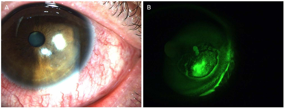
Figure 1 Slit-lamp photography of a patient who was diagnosed with recurrent corneal erosion. A 60-year-old male patient presented with a ragged and greyish appearance of the epithelium in the left eye (A) and abnormal fluorescein staining (B).
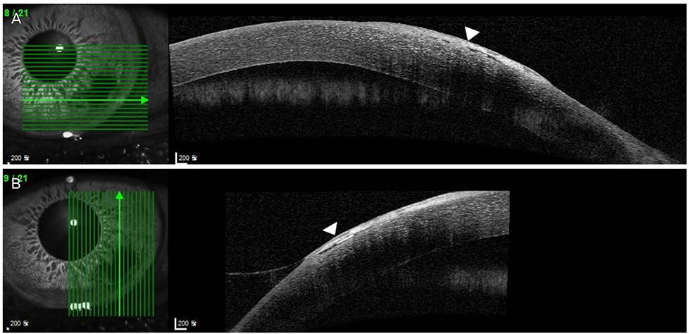
Figure 2 Anterior segment optical coherence tomography of the left cornea. Hyper-reflective signals pointed by arrowheads involving both Bowman's layer and superficial cornea (A, B).
Reference
-
1. Kirkwood BJ. Recurrent corneal erosion: characteristics and management options. Insight. 2007; 32:14–17.2. Ramamurthi S, Rahman MQ, Dutton GN, Ramaesh K. Pathogenesis, clinical features and management of recurrent corneal erosions. Eye (Lond). 2006; 20:635–644.
Article3. Tripathi RC, Bron AJ. Ultrastructural study of non-traumatic recurrent corneal erosion. Br J Ophthalmol. 1972; 56:73–85.
Article4. Sakimoto T, Sawa M. Metalloproteinases in corneal diseases: degradation and processing. Cornea. 2012; 31:Suppl 1. S50–S56.5. Chen YT, Huang CW, Huang FC, et al. The cleavage plane of corneal epithelial adhesion complex in traumatic recurrent corneal erosion. Mol Vis. 2006; 12:196–204.6. Goldman JN, Dohlman CH, Kravitt BA. The basement membrane of the human cornea in recurrent epithelial erosion syndrome. Trans Am Acad Ophthalmol Otolaryngol. 1969; 73:471–481.7. Wood TO. Recurrent erosion. Trans Am Ophthalmol Soc. 1984; 82:850–898.8. Watson S, Lee H. Interventions for recurrent corneal erosion: a cochrane systematic review. Eye (Lond). 2013; 27:1330–1331.
Article9. Dursun D, Kim MC, Solomon A, Pflugfelder SC. Treatment of recalcitrant recurrent corneal erosions with inhibitors of matrix metalloproteinase-9, doxycycline and corticosteroids. Am J Ophthalmol. 2001; 132:8–13.
Article10. Das S, Seitz B. Recurrent corneal erosion syndrome. Surv Ophthalmol. 2008; 53:3–15.
Article11. Singh RP, Raj D, Pherwani A, et al. Alcohol delamination of the corneal epithelium for recalcitrant recurrent corneal erosion syndrome: a prospective study of efficacy and safety. Br J Ophthalmol. 2007; 91:908–911.
Article12. Soong HK, Farjo Q, Meyer RF, Sugar A. Diamond burr superficial keratectomy for recurrent corneal erosions. Br J Ophthalmol. 2002; 86:296–298.
Article13. McLean EN, MacRae SM, Rich LF. Recurrent erosion. Treatment by anterior stromal puncture. Ophthalmology. 1986; 93:784–788.14. Ohman L, Fagerholm P. The influence of excimer laser ablation on recurrent corneal erosions: a prospective randomized study. Cornea. 1998; 17:349–352.15. Williams R, Buckley RJ. Pathogenesis and treatment of recurrent erosion. Br J Ophthalmol. 1985; 69:435–437.
Article16. Fraunfelder FW, Cabezas M. Treatment of recurrent corneal erosion by extended-wear bandage contact lens. Cornea. 2011; 30:164–166.
Article17. Kenyon KR, Fogle JA, Stone DL, Stark WJ. Regeneration of corneal epithelial basement membrane following thermal cauterization. Invest Ophthalmol Vis Sci. 1977; 16:292–301.18. Mandić Z, Bednar I, Šarić D. Modern approach in the treatment of recurrent corneal erosion. Acta Clin Croatica. 2007; 46:Suppl 1. 25–30.19. Hykin PG, Foss AE, Pavesio C, Dart JK. The natural history and management of recurrent corneal erosion: a prospective randomized trial. Eye (Lond). 1994; 8(Pt 1):35–40.20. Hsu JK, Rubinfeld RS, Barry P, Jester JV. Anterior stromal puncture. Immunohistochemical studies in human corneas. Arch Ophthalmol. 1993; 111:1057–1063.21. Lee SW, Choi TH. Anterior stromal puncture with 26-gauge needle for recurrent corneal erosion: a report of five cases. J Korean Ophthalmol Soc. 2003; 44:511–516.22. Rubinfeld RS, Laibson PR, Cohen EJ, et al. Anterior stromal puncture for recurrent erosion: further experience and new instrumentation. Ophthalmic Surg. 1990; 21:318–326.
Article23. Choi M, Jung JW, Seo KY, et al. Comparison of Nd:YAG laser versus conservative management in the treatment of recurrent corneal erosion. J Korean Ophthalmol Soc. 2015; 56:687–693.
Article24. Kim SY, Ko BY. Evaluation of anterior stromal puncture using Nd:YAG laser for refractory recurrent corneal erosion. J Korean Ophthalmol Soc. 2015; 56:331–338.
Article25. O'Brart DP, Muir MG, Marshall J. Phototherapeutic keratectomy for recurrent corneal erosions. Eye (Lond). 1994; 8(Pt 4):378–383.26. Ko BY, Lee GW. Clinical results of phototherapeutic keratectomy for refractory recurrent corneal erosion. J Korean Ophthalmol Soc. 2011; 52:392–400.
Article27. Baryla J, Pan YI, Hodge WG. Long-term efficacy of phototherapeutic keratectomy on recurrent corneal erosion syndrome. Cornea. 2006; 25:1150–1152.
Article28. Bae KH, Ahn M, Cho NC, You IC. Clinical presentation and treatment outcomes of recurrent corneal erosion. J Korean Ophthalmol Soc. 2016; 57:555–561.
Article29. Brown N, Bron A. Recurrent erosion of the cornea. Br J Ophthalmol. 1976; 60:84–96.
Article30. Shin DY, Chung SH. Efficacy of anterior stromal puncture using 5% NaCl eye drops for prolonged time in recurrent corneal erosion syndrome. J Korean Ophthalmol Soc. 2017; 58:503–508.
Article31. Liu C, Buckley R. The role of the therapeutic contact lens in the management of recurrent corneal erosions: a review of treatment strategies. CLAO J. 1996; 22:79–82.32. Ahad MA, Anandan M, Tah V, et al. Randomized controlled study of ocular lubrication versus bandage contact lens in the primary treatment of recurrent corneal erosion syndrome. Cornea. 2013; 32:1311–1314.
Article33. Lee SE, Kim SR, Park MJ. Oxygen permeability of soft contact lenses in different pH, osmolality and buffering solution. Int J Ophthalmol. 2015; 8:1037–1042.34. Aiello JP, Insler MS. The effects of hypotonic and hypertonic solutions on the fluid content of hydrophilic contact lenses. Am J Ophthalmol. 1985; 99:521–523.
Article35. Stahl U, Wilcox M, Stapleton F. Role of hypo-osmotic saline drops in ocular comfort during contact lens wear. Cont Lens Anterior Eye. 2010; 33:68–75.
Article36. Kanpolat A, Uçakhan OO. Therapeutic use of focus night & day contact lense. Cornea. 2003; 22:726–734.37. Ambroziak AM, Szaflik JP, Szaflik J. Therapeutic use of silicone hydrogel contact lens in selected clinical cases. Eye Contact Lens. 2004; 30:63–67.38. Green M, Apel A, Stapleton F. Risk factors and causative organisms in microbial keratitis. Cornea. 2008; 27:22–27.
Article39. Stern GA. Contact lens associated bacterial keratitis: past, present, and future. CLAO J. 1998; 24:52–56.40. Hahn YH, Hahn TW, Choi SH, et al. Epidemiology of infectious keratitis [I] a multi-center study. J Korean Ophthalmol Soc. 1998; 39:1633–1651.41. Stapleton F, Keay L, Edwards K, et al. The incidence of contact lens-related microbial keratitis in Australia. Ophthalmology. 2008; 115:1655–1662.
Article42. Cho CH, Lee SB. Analysis of inpatients with contact lens related bacterial keratitis:causative microorganisms, clinical aspects, and prognostic factors. J Korean Ophthalmol Soc. 2013; 54:1327–1338.43. Brem H, Tomic-Canic M. Cellular and molecular basis of wound healing in diabetes. J Clin Invest. 2007; 117:1219–1222.
Article44. Liu ZJ, Velazquez OC. Hyperoxia, endothelial progenitor cell mobilization, and diabetic wound healing. Antioxid Redox Signal. 2008; 10:1869–1882.
Article45. Sun H, Mi X, Gao N, et al. Hyperglycemia-suppressed expression of Serpine1 contributes to delayed epithelial wound healing in diabetic mouse corneas. Invest Ophthalmol Vis Sci. 2015; 56:3383–3392.
Article
- Full Text Links
-
- Actions
-
Cited
- CITED
-
- Close
- Share
-
- Similar articles
-
- Clinical Features of Corneal Erosion in Contact Lens Wearers
- Clinical Study of Eye-Me(R) Soft Contact lens
- Corneal Complications in Contact Lens Wearer
- Complications of Contact Lens Wear After Radial Keratotomy in an Animal Model
- Clinical Evaluation of Bausch and Lomb O3/O4 Soft Contact Lenses for Extended Wear
