A Rare Manifestation of Solitary Primary Bone Lymphoma of the Finger: a Case Report
- Affiliations
-
- 1Department of Radiology, Pusan National University Hospital, Busan, Korea. yssongrad@gmail.com
- 2Department of Radiology, Pusan National University Yangsan Hospital, Busan, Korea.
- 3Department of Pathology, Pusan National University Hospital, Busan, Korea.
- 4Department of Orthopedic Surgery, Pusan National University Hospital, Busan, Korea.
- KMID: 2431108
- DOI: http://doi.org/10.13104/imri.2018.22.4.240
Abstract
- Primary extranodal bone lymphoma involving the peripheral extremities is extremely rare. Here, we report a definitive case of diffuse large B-cell lymphoma involving the phalangeal bone of the 3rd finger. Systemic evaluation revealed the lesion as the only site of lymphoma involvement.
Keyword
MeSH Terms
Figure
-
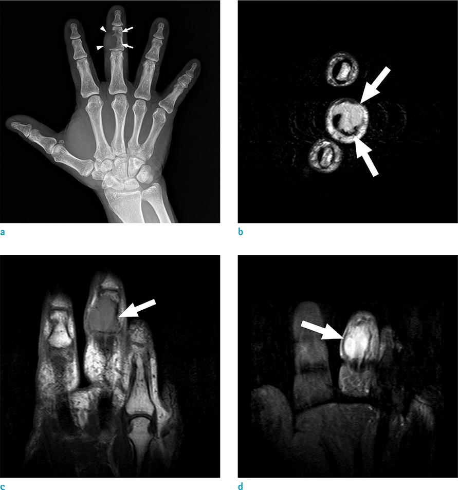
Fig. 1 A 71-year-old-man with DLBCL involving 3rd finger. (a) Plain radiograph shows osteolytic bony mass (arrows) with soft-tissue component (arrowheads) involving right third middle phalanx. (b) Axial T2-weighted image reveals a high-signal intensity mass (arrows) associated with the right third middle phalanx. The mass originated in the intramedullary cavity constituting the large soft-tissue component, abutting the flexor tendon of the third finger. (c) Coronal T1-weighted image indicates a mass of intermediate signal intensity (arrow). (d) Coronal T1-weighted image with fat suppression after gadolinium administration shows homogenous enhancement of the mass (arrow).
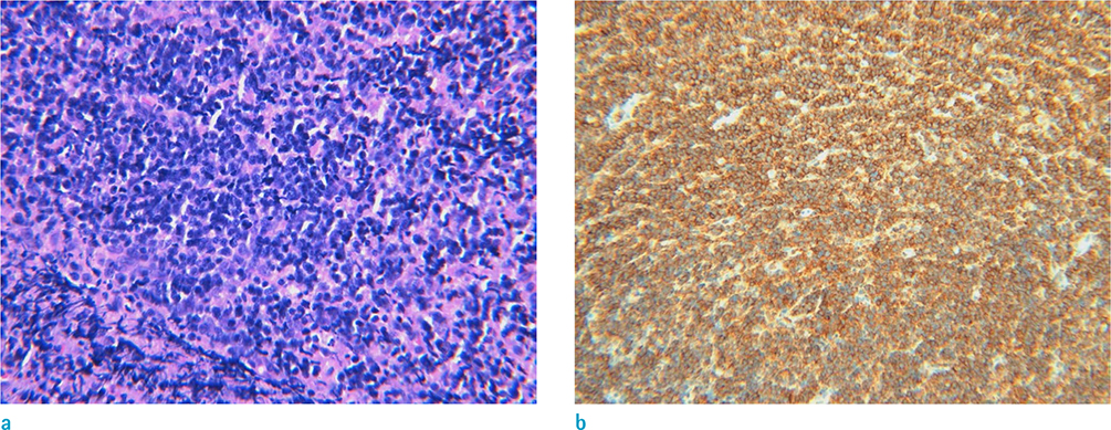
Fig. 2 Microscopic findings of the finger mass. (a) Microscopic finding (Hematoxylin & Eosin stain, × 200) shows diffuse infiltration of atypical cells. (b) Immunohistochemical staining was positive for leukocyte common antigen.
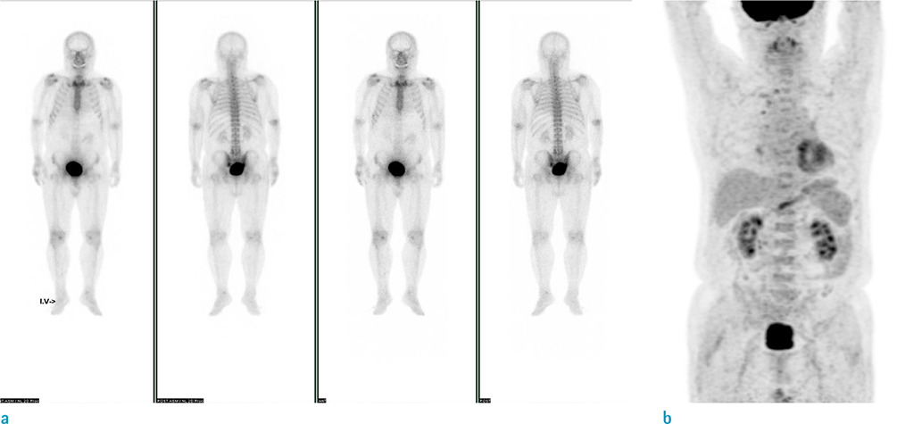
Fig. 3 Systemic evaluation of other organ involvement. (a) Bone scan reveals no evidence of other bone involvement. (b) PET-CT reveals no evidence of other organ involvement of lymphoma.
Reference
-
1. Krishnan A, Shirkhoda A, Tehranzadeh J, Armin AR, Irwin R, Les K. Primary bone lymphoma: radiographic-MR imaging correlation. Radiographics. 2003; 23:1371–1383. discussion 1384-1387.
Article2. Pobirci O, Rosca E, Pobirci DD, Nichita A. Primary diffuse large B-cell lymphoma of the humerus. Case report and review of literature. Med Con. 2014; 9:69–72.3. Mulligan ME, McRae GA, Murphey MD. Imaging features of primary lymphoma of bone. AJR Am J Roentgenol. 1999; 173:1691–1697.
Article4. de Camargo OP, dos Santos Machado TM, Croci AT, et al. Primary bone lymphoma in 24 patients treated between 1955 and 1999. Clin Orthop Relat Res. 2002; 271–280.
Article5. Vincent JM, Ng YY, Norton AJ, Armstrong P. Case report: primary lymphoma of bone--MRI appearances with pathological correlation. Clin Radiol. 1992; 45:407–409.6. Jawad MU, Schneiderbauer MM, Min ES, Cheung MC, Koniaris LG, Scully SP. Primary lymphoma of bone in adult patients. Cancer. 2010; 116:871–879.
Article7. Galati V, Wortmann F, Stang FH, Thorns C, Mailander P, Kisch T. A rare manifestation of primary bone lymphoma: solitary diffuse large B-cell lymphoma of the little finger. J Hand Surg Am. 2018; 43:779.e1–779.e4.
Article8. Coley BL, Higinbotham NL, Groesbeck HP. Primary reticulum-cell sarcoma of bone; summary of 37 cases. Radiology. 1950; 55:641–658.9. Li X, Xu-Monette ZY, Yi S, et al. Primary bone lymphoma exhibits a favorable prognosis and distinct gene expression signatures resembling diffuse large B-cell lymphoma derived from centrocytes in the germinal center. Am J Surg Pathol. 2017; 41:1309–1321.
Article10. Phillips WC, Kattapuram SV, Doseretz DE, et al. Primary lymphoma of bone: relationship of radiographic appearance and prognosis. Radiology. 1982; 144:285–290.
Article
- Full Text Links
-
- Actions
-
Cited
- CITED
-
- Close
- Share
-
- Similar articles
-
- A Case of Solitary Neurofibroma on the Finger with Nail Deformity
- Disseminated Primary Non-Hodgkin's Lymphoma of Bone: A Case Report
- Sonographic Findings of Primary Tracheal Lymphoma: A Case Report
- Primary Cutaneous Monomorphous Lymphoma: A Report of 3 Cases
- Primary Malignant Lymphoma of Lung: A Case Report
