Korean Circ J.
2019 Jan;49(1):113-114. 10.4070/kcj.2018.0243.
Ovoid-shaped Left Main Coronary Calcified Aneurysm Leading to Unstable Angina Requiring Surgical Intervention
- Affiliations
-
- 1Division of Cardiology, Department of Internal Medicine, Dongguk University College of Medicine and Dongguk University Medical Center, Goyang, Korea. rsy008@gmail.com
- KMID: 2430755
- DOI: http://doi.org/10.4070/kcj.2018.0243
Abstract
- No abstract available.
MeSH Terms
Figure
-
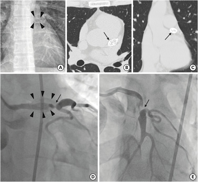
Figure 1 An ovoid mass near left side of the aorta in chest X-ray (arrow heads) (A). A condensed calcified mass located on LMCA (arrow) in horizontal section (B) and coronal section (C) of chest CT. Diagnostic CAG demonstrated a severe stenosis of distal portion of LMCA (arrows) surrounded by the calcified mass (arrow heads) on RAO (D) and LAO (E) view.LMCA = left main coronary artery; CT = computed tomography; CAG = coronary angiography; RAO = right anterior oblique; LAO = left anterior oblique.
Reference
-
1. Aggarwal A, Srivastava S, Velmurugan M. Newer perspectives of coronary artery disease in young. World J Cardiol. 2016; 8:728–734. PMID: 28070240.2. Beiser AS, Takahashi M, Baker AL, Sundel RP, Newburger JW. A predictive instrument for coronary artery aneurysms in Kawasaki disease. US Multicenter Kawasaki Disease Study Group. Am J Cardiol. 1998; 81:1116–1120. PMID: 9605052.3. Bang JS, Kim GB, Kwon BS, et al. Long-term prognosis for patients with Kawasaki disease complicated by large coronary aneurysm (diameter ≥6 mm). Korean Circ J. 2017; 47:516–522. PMID: 28765744.
- Full Text Links
-
- Actions
-
Cited
- CITED
-
- Close
- Share
-
- Similar articles
-
- A Case of Left Coronary Artery Milking Treated by Direct Stenting During Percutaneous Coronary Intervention in A Patient with Unstable Angina
- A Cses of Total Occlusion of the Left Main Coronary Artery
- Unprotected Left Main Percutaneous Coronary Intervention in a 108-Year-Old Patient
- CABG for Treating Unstable Angina with Multivessel Coronary Artery Aneurysms: A case report
- The Level of Interleukin-6 in Coronary sinus and peripheral blood in patiens with unstable angina after coronary intervention
