Schematic of mean thickness distribution on the lateral aspect of the canine frontal sinus as an experimental model of sinus surgery
- Affiliations
-
- 1Department of Dental Hygiene, Division of Health Science, Dongseo University, Busan, Korea.
- 2Division in Anatomy and Developmental Biology, Department of Oral Biology, Yonsei University College of Dentistry, Seoul, Korea. hks318@yuhs.ac
- 3Department of Occupational Therapy, College of Health and Welfare, Semyung University, Jecheon, Korea.
- 4Department of Periodontology, Research Institute for Periodontal Regeneration, Yonsei University College of Dentistry, Seoul, Korea.
- KMID: 2430190
- DOI: http://doi.org/10.5115/acb.2018.51.4.236
Abstract
- The dog frontal sinus may represent an alternative model dental implant research; its topographical resemblance to the maxillary sinus renders it a potentially favorable experimental environment. The aim of this study was thus to elucidate the anatomical configuration of the canine frontal sinus and histological characteristics, and to determine whether it could be a new canine experimental model for dental implant research. Twenty-four sides of canine frontal bones were harvested. The distance from the nasion to the emerging point of the lateral aspect of the canine frontal sinus was measured with the aid of Lucion software. The thicknesses of the canine frontal sinus wall were measured, and the two specimens stained with hematoxylin and eosin. The mean distance from the nasion to the emerging point of the lateral aspect of the canine frontal sinus was 16.0 mm. The mean thicknesses of the canine frontal bone at 3, 6, 9, 12, and 15 mm lateral to the midsagittal plane were 2.3, 2.7, 3.2, 3.8, and 3.7 mm, respectively. The canine frontal sinus was lined with pseudostratified ciliated columnar epithelium. These data suggest that the canine frontal sinus is a suitable alternative to the canine maxillary sinus as a model for studying various sinus augmentation protocols.
Keyword
MeSH Terms
Figure
-
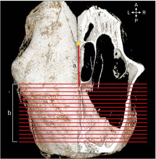
Fig. 1 Parameters measured in the canine frontal bone and sinus with the aid of Lucion software. a, the distance from nasion (yellow asterisk) to the emerging point of the lateral aspect of the canine frontal sinus; b, the 16 coronal sections imaged from the emerging point of the lateral aspect of the canine frontal sinus in the posterior direction at 1-mm intervals. A, anterior; P, posterior; L, left; R, right.

Fig. 2 The thickness of the canine frontal bone was measured at 3, 6, 9, 12, and 15 mm lateral to the midsagittal plane (double-headed arrows) at 3-mm intervals on each coronally sectioned image (CS1–CS16). A, anterior; P, posterior; L, left; R, right.
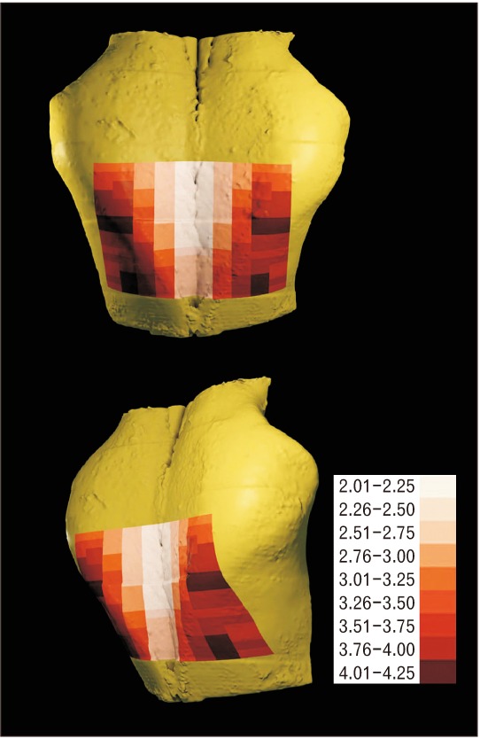
Fig. 3 The thickness of the canine frontal sinus wall (mm).
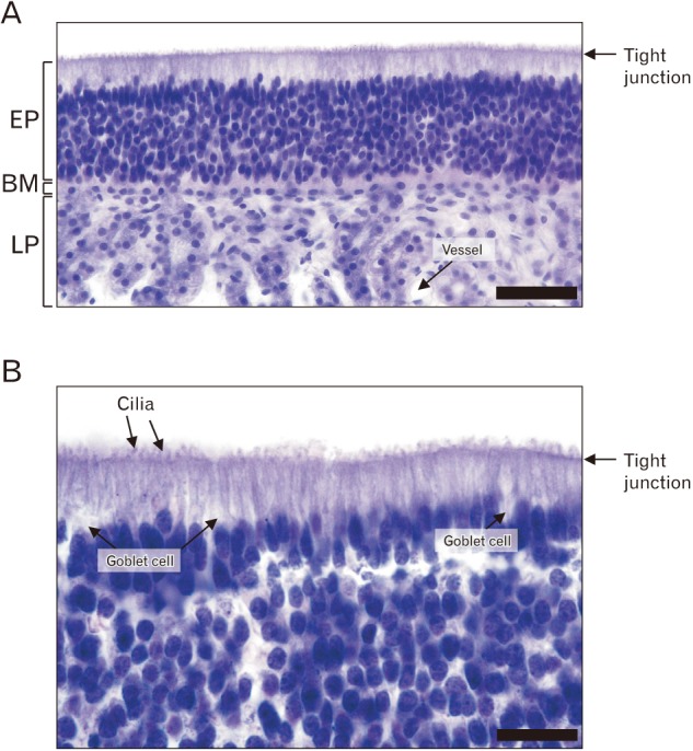
Fig. 4 Histological examination of the canine frontal sinus mucosa (A, ×400; B, ×1,000). Scale bars=50 µm (A), 200 µm (B). EP, epithelium; BM, basement membrane; LP, lamina propria.
Reference
-
1. Mayfield LJ, Skoglund A, Hising P, Lang NP, Attström R. Evaluation following functional loading of titanium fixtures placed in ridges augmented by deproteinized bone mineral: a human case study. Clin Oral Implants Res. 2001; 12:508–514. PMID: 11564112.2. Soncini M, Rodriguez y, Pietrabissa R, Quaglini V, Rizzo S, Zaffe D. Experimental procedure for the evaluation of the mechanical properties of the bone surrounding dental implants. Biomaterials. 2002; 23:9–17. PMID: 11762859.3. Bertin PM. The use of multiple implant modalities in the management of the edentulous patient. Compendium. 1989; 10:657–658. 660–661. PMID: 2700681.4. Bishop K, Addy L, Knox J. Modern restorative management of patients with congenitally missing teeth: 4. The role of implants. Dent Update. 2007; 34:79–80. 82–84. PMID: 17432771.5. Misch CE. Maxillary sinus augmentation for endosteal implants: organized alternative treatment plans. Int J Oral Implantol. 1987; 4:49–58. PMID: 3269837.6. van den Bergh JP, ten Bruggenkate CM, Krekeler G, Tuinzing DB. Sinusfloor elevation and grafting with autogenous iliac crest bone. Clin Oral Implants Res. 1998; 9:429–435. PMID: 11429944.7. Kahnberg KE, Ekestubbe A, Gröndahl K, Nilsson P, Hirsch JM. Sinus lifting procedure. I. One-stage surgery with bone transplant and implants. Clin Oral Implants Res. 2001; 12:479–487. PMID: 11564108.8. Lee SH, Choi BH, Li J, Jeong SM, Kim HS, Ko CY. Comparison of corticocancellous block and particulate bone grafts in maxillary sinus floor augmentation for bone healing around dental implants. Oral Surg Oral Med Oral Pathol Oral Radiol Endod. 2007; 104:324–328. PMID: 17428698.9. Brumund KT, Graham SM, Beck KC, Hoffman EA, McLennan G. The effect of maxillary sinus antrostomy size on xenon ventilation in the sheep model. Otolaryngol Head Neck Surg. 2004; 131:528–533. PMID: 15467631.10. Asai S, Shimizu Y, Ooya K. Maxillary sinus augmentation model in rabbits: effect of occluded nasal ostium on new bone formation. Clin Oral Implants Res. 2002; 13:405–409. PMID: 12175378.11. Bravetti P, Membre H, Marchal L, Jankowski R. Histologic changes in the sinus membrane after maxillary sinus augmentation in goats. J Oral Maxillofac Surg. 1998; 56:1170–1176. PMID: 9766543.12. Hanisch O, Tatakis DN, Rohrer MD, Wöhrle PS, Wozney JM, Wikesjö UM. Bone formation and osseointegration stimulated by rhBMP-2 following subantral augmentation procedures in nonhuman primates. Int J Oral Maxillofac Implants. 1997; 12:785–792. PMID: 9425759.13. Haas R, Donath K, Födinger M, Watzek G. Bovine hydroxyapatite for maxillary sinus grafting: comparative histomorphometric findings in sheep. Clin Oral Implants Res. 1998; 9:107–116. PMID: 9663038.14. Watanabe K, Niimi A, Ueda M. Autogenous bone grafts in the rabbit maxillary sinus. Oral Surg Oral Med Oral Pathol Oral Radiol Endod. 1999; 88:26–32. PMID: 10442941.15. Liu N, Sun F, Xu C, Lin T, Lu E. A comparative study of dog models for osteotome sinus floor elevation and dental implants in posterior maxilla subjacent to the maxillary sinus. Oral Surg Oral Med Oral Pathol Oral Radiol. 2013; 115:e15–e20. PMID: 22749358.16. Jung JH, Choi BH, Zhu SJ, Lee SH, Huh JY, You TM, Lee HJ, Li J. The effects of exposing dental implants to the maxillary sinus cavity on sinus complications. Oral Surg Oral Med Oral Pathol Oral Radiol Endod. 2006; 102:602–605. PMID: 17052635.17. Toyohiko H, Takao W, Junichi S. An experimental study on maxillary sinus floor elevation and simultaneous implant placement into the frontal sinus of dogs using bone substitutes. Tsurumi Shigaku. 2005; 31:65–80.18. Miller ME, Christensen GC, Evans HE. Anatomy of the dog. Philadelphia, PA: W. B. Saunders;1964.19. Estaca E, Cabezas J, Usón J, Sánchez-Margallo F, Morell E, Latorre R. Maxillary sinus-floor elevation: an animal model. Clin Oral Implants Res. 2008; 19:1044–1048. PMID: 18828821.20. Bretan O, Nogueira EA, Silva EF, Trindade SH. Training the osteoplastic flap technique in dogs. Braz J Otorhinolaryngol. 2005; 71:140–144. PMID: 16446908.21. Burrow R, McCarroll D, Baker M, Darby P, McConnell F, Cripps P. Frontal sinus depth at four landmarks in breeds of dog typically affected by sinonasal aspergillosis. Vet Rec. 2012; 170:20. PMID: 22016511.22. Esfahanizadeh N, Rokn AR, Paknejad M, Motahari P, Daneshparvar H, Shamshiri A. Comparison of lateral window and osteotome techniques in sinus augmentation: histological and histomorphometric evaluation. J Dent (Tehran). 2012; 9:237–246. PMID: 23119133.23. Kolerman R, Moses O, Artzi Z, Barnea E, Tal H. Maxillary sinus augmentation by the crestal core elevation technique. J Periodontol. 2011; 82:41–51. PMID: 20731587.24. Del Fabbro M, Corbella S, Weinstein T, Ceresoli V, Taschieri S. Implant survival rates after osteotome-mediated maxillary sinus augmentation: a systematic review. Clin Implant Dent Relat Res. 2012; 14(Suppl 1):e159–e168. PMID: 22082056.25. Yang HM, Bae HE, Won SY, Hu KS, Song WC, Paik DJ, Kim HJ. The buccofacial wall of maxillary sinus: an anatomical consideration for sinus augmentation. Clin Implant Dent Relat Res. 2009; 11(Suppl 1):e2–e6. PMID: 19438968.26. van den Bergh JP, ten Bruggenkate CM, Disch FJ, Tuinzing DB. Anatomical aspects of sinus floor elevations. Clin Oral Implants Res. 2000; 11:256–265. PMID: 11168217.27. Peleg M, Garg AK, Mazor Z. Predictability of simultaneous implant placement in the severely atrophic posterior maxilla: a 9-year longitudinal experience study of 2132 implants placed into 731 human sinus grafts. Int J Oral Maxillofac Implants. 2006; 21:94–102. PMID: 16519187.28. Fugazzotto PA, Vlassis J. Report of 1633 implants in 814 augmented sinus areas in function for up to 180 months. Implant Dent. 2007; 16:369–378. PMID: 18091165.29. Beretta M, Cicciù M, Bramanti E, Maiorana C. Schneider membrane elevation in presence of sinus septa: anatomic features and surgical management. Int J Dent. 2012; 2012:261905. PMID: 22848223.30. Betts NJ, Miloro M. Modification of the sinus lift procedure for septa in the maxillary antrum. J Oral Maxillofac Surg. 1994; 52:332–333. PMID: 8308638.31. Tatum H Jr. Maxillary and sinus implant reconstructions. Dent Clin North Am. 1986; 30:207–229. PMID: 3516738.32. Aimetti M, Massei G, Morra M, Cardesi E, Romano F. Correlation between gingival phenotype and Schneiderian membrane thickness. Int J Oral Maxillofac Implants. 2008; 23:1128–1132. PMID: 19216284.33. Pommer B, Unger E, Suto D, Hack N, Watzek G. Mechanical properties of the Schneiderian membrane in vitro. Clin Oral Implants Res. 2009; 20:633–637. PMID: 19281504.34. Tos M, Mogensen C. Mucus production in the nasal sinuses. Acta Otolaryngol Suppl. 1979; 360:131–134. PMID: 287326.35. Sul SH, Choi BH, Li J, Jeong SM, Xuan F. Histologic changes in the maxillary sinus membrane after sinus membrane elevation and the simultaneous insertion of dental implants without the use of grafting materials. Oral Surg Oral Med Oral Pathol Oral Radiol Endod. 2008; 105:e1–e5.36. Choi BH, Zhu SJ, Jung JH, Lee SH, Huh JY. The use of autologous fibrin glue for closing sinus membrane perforations during sinus lifts. Oral Surg Oral Med Oral Pathol Oral Radiol Endod. 2006; 101:150–154. PMID: 16448914.37. Abramson AL, Eason RL. Experimental frontal sinus obliteration: long-term results following removal of the mucous membrane lining. Laryngoscope. 1977; 87:1066–1073. PMID: 875567.
- Full Text Links
-
- Actions
-
Cited
- CITED
-
- Close
- Share
-
- Similar articles
-
- A Case of Endoscopic Sinus Surgery for Lateral Frontal Sinusitis
- Frontal Sinus Stenting in Endoscopic Sinus Surgery
- Endoscopic Frontal Sinus Surgery
- A study on the mandibular growth prediction and size of the frontal sinus
- A Case of Frontal Mococele Treated with Transblepharoplasty Approach Combined with Endoscopic Approach
