J Cardiovasc Imaging.
2018 Dec;26(4):253-255. 10.4250/jcvi.2018.26.e23.
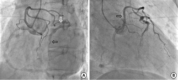
Three in One Coronary Pathology: Finding the Culprit
- Affiliations
-
- 1Department of Cardiology, Sanjay Gandhi Postgraduate, Institute of Medical Sciences, Lucknow, India. drroopalik@gmail.com
- 2Department of Radiology, Sanjay Gandhi Postgraduate, Institute of Medical Sciences, Lucknow, India.
- KMID: 2430002
- DOI: http://doi.org/10.4250/jcvi.2018.26.e23
Abstract
- No abstract available.
MeSH Terms
Figure
Reference
-
1. Angelini P. Coronary artery anomalies: an entity in search of an identity. Circulation. 2007; 115:1296–1305.2. Pursnani A, Jacobs JE, Saremi F, et al. Coronary CTA assessment of coronary anomalies. J Cardiovasc Comput Tomogr. 2012; 6:48–59.
Article
- Full Text Links
- Actions
-
Cited
- CITED
-
- Close
- Share
- Similar articles
-
- Treat or Not to Treat Non-culprit Coronary Artery with Significant Stenosis during Primary Percutaneous Coronary Intervention
- Physiologic Evaluation of Microvascular Damage in Culprit Vessel After Successful Primary Percutaneous Coronary Intervention for ST-elevation Myocardial Infarction Patients
- Missing Right Coronary Artery in a Patient with Acute Inferior ST Segment Elevation Myocardial Infarction: A Case of Extremely Rare Variation of Coronary Anatomy
- Accuracy of the Electrocardiographic Criteria for Predicting the Right or Left Circumflex Coronary Artery as the Culprit Coronary Artery in Acute Inferior Myocardial Infarction
- ST Segment Depression in Lateral Leads in Inferior Wall Acute Myocardial Infarction


