Semiautomatic Three-Dimensional Threshold-Based Cardiac Computed Tomography Ventricular Volumetry in Repaired Tetralogy of Fallot: Comparison with Cardiac Magnetic Resonance Imaging
- Affiliations
-
- 1Department of Radiology and Research Institute of Radiology, University of Ulsan College of Medicine, Asan Medical Center, Seoul, Korea. ghw68@hanmail.net
- KMID: 2429924
- DOI: http://doi.org/10.3348/kjr.2018.0237
Abstract
OBJECTIVE
To assess the accuracy and potential bias of computed tomography (CT) ventricular volumetry using semiautomatic three-dimensional (3D) threshold-based segmentation in repaired tetralogy of Fallot, and to compare them to those of two-dimensional (2D) magnetic resonance imaging (MRI).
MATERIALS AND METHODS
This retrospective study evaluated 32 patients with repaired tetralogy of Fallot who had undergone both cardiac CT and MRI within 3 years. For ventricular volumetry, semiautomatic 3D threshold-based segmentation was used in CT, while a manual simplified contouring 2D method was used in MRI. The indexed ventricular volumes were compared between CT and MRI. The indexed ventricular stroke volumes were compared with the indexed arterial stroke volumes measured using phase-contrast MRI. The mean differences and degrees of agreement in the indexed ventricular and stroke volumes were evaluated using Bland-Altman analysis.
RESULTS
The indexed end-systolic (ES) volumes showed no significant difference between CT and MRI (p > 0.05), while the indexed end-diastolic (ED) volumes were significantly larger on CT than on MRI (93.6 ± 17.5 mL/m² vs. 87.3 ± 15.5 mL/m² for the left ventricle [p < 0.001] and 177.2 ± 39.5 mL/m² vs. 161.7 ± 33.1 mL/m² for the right ventricle [p < 0.001], respectively). The mean differences between CT and MRI were smaller for the indexed ES volumes (2.0-2.5 mL/m²) than for the indexed ED volumes (6.3-15.5 mL/m²). CT overestimated the stroke volumes by 14-16%. With phase-contrast MRI as a reference, CT (7.2-14.3 mL/m²) showed greater mean differences in the indexed stroke volumes than did MRI (0.8-3.3 mL/m²; p < 0.005).
CONCLUSION
Compared to 2D MRI, CT ventricular volumetry using semiautomatic 3D threshold-based segmentation provides comparable ES volumes, but overestimates the ED and stroke volumes in patients with repaired tetralogy of Fallot.
Keyword
MeSH Terms
Figure
-
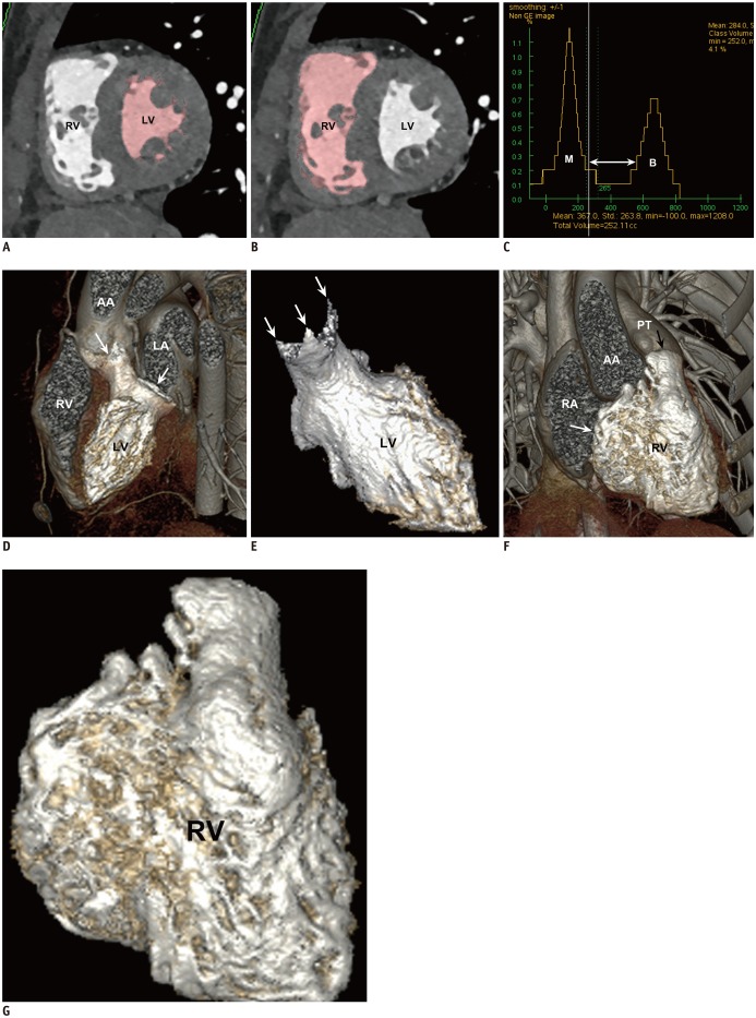
Fig. 1 18-year-old female patient with repaired tetralogy of Fallot.Short-axis reformatted ES cardiac computed tomographic images show left ventricular cavity (A) and right ventricular cavity (B) segmented using three-dimensional threshold-based method in pink. Papillary muscles and trabeculations are largely excluded from ventricular cavity by using this method. C. Histogram shows that threshold (vertical line) used for segmentation is more closely located to distribution curve of M than to that of ventricular B in order to exclude voxels consisting of 100% ventricular M consistently. As result, small subset of voxels comprising variable mixtures of myocardial and B tissues between two peaks (horizontal arrow) is included in ventricular cavity. D. Oblique sagittal volume-rendered computed tomographic image demonstrates valve planes (arrows) of highlighted left ventricular cavity after manual segmentation. E. Resultant left ventricular ES volume is approximately 49.3 mL. Three commissures (arrows) of aortic valve are clearly noted. F. Oblique coronal volume-rendered computed tomographic image displays valve planes (arrows) of highlighted right ventricular cavity after manual segmentation. AA = ascending aorta, B = blood, ES = end-systolic, LA = left atrium, LV = left ventricle, M = myocardium, PT = pulmonary trunk, RA = right atrium, RV = right ventricle. G. Resultant right ventricular ES volume is 97.6 mL. ES = end-systolic, RV = right ventricle
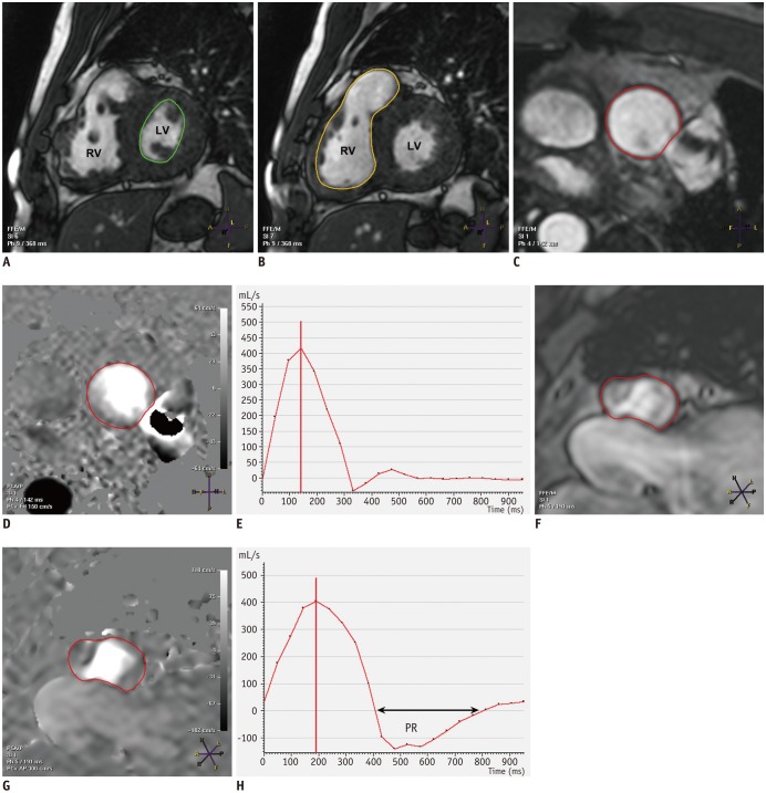
Fig. 2 36-year-old male patient with repaired tetralogy of Fallot and left pulmonary artery stent placement.A, B. Two-dimensional short-axis ES cine MR images show manually traced left ventricular cavity (green line in A) and right ventricular cavity (yellow line in B) acquired using simplified contouring method. Papillary muscles and trabeculations are included in ventricular cavity in this method. Magnitude (C) and phase (D) images of through-plane phase-contrast MRI for ascending aorta show semiautomatically drawn vessel outline in red. E. Ascending aortic flow curve is then automatically generated throughout cardiac cycle. Magnitude (F) and phase (G) images of through-plane phase-contrast MRI for pulmonary trunk show semiautomatically drawn vessel outline in red. H. Pulmonary arterial flow curve is then automatically generated throughout cardiac cycle. PR fraction is noted as area under baseline (horizontal arrow). MR = magnetic resonance, MRI = magnetic resonance imaging, PR = pulmonary regurgitation
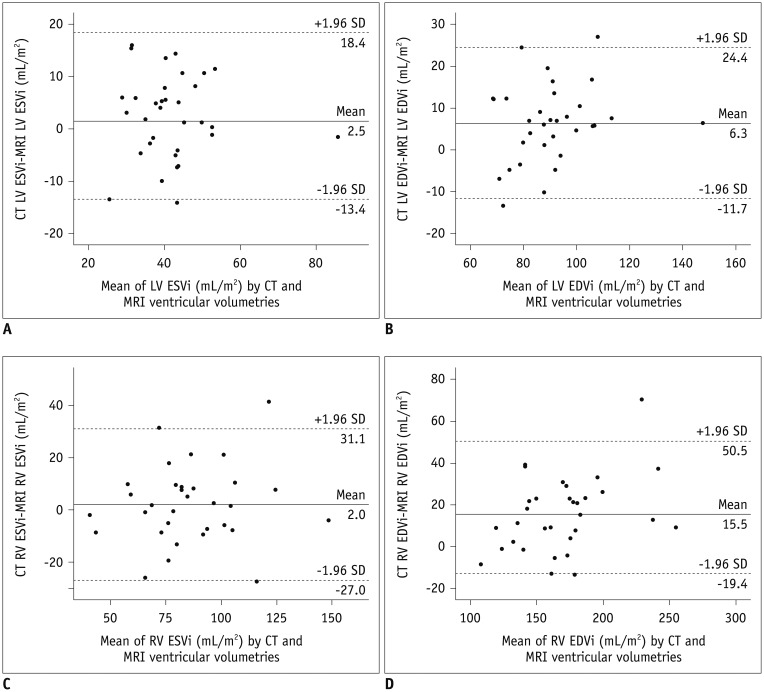
Fig. 3 Bland-Altman analysis between ventricular volumes measured using cardiac CT and short-axis cine MRI.A–D. Bland-Altman plots illustrate average bias and degree of agreement in indexed left ventricular ES (A) and ED (B) volumes as well as between indexed right ventricular ES (C) and ED (D) volumes for two methods. CT = computed tomography, ED = end-diastolic, EDVi = indexed end-diastolic volume, ESVi = indexed end-systolic volume, SD = standard deviation
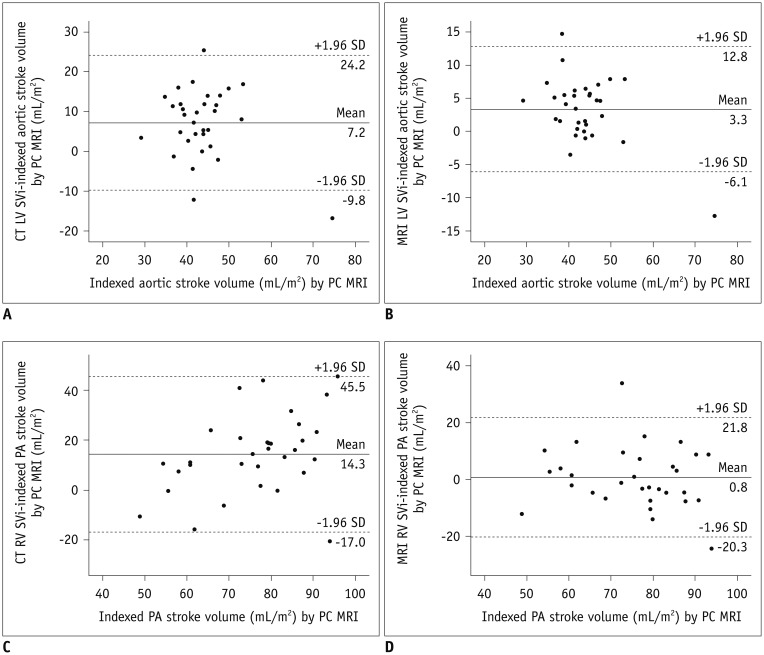
Fig. 4 Bland-Altman analysis between indexed arterial stroke volumes measured using phase-contrast MRI and indexed ventricular stroke volumes.Bland-Altman plots illustrate average bias and degree of agreement in indexed left ventricular stroke volumes between phase-contrast MRI and CT ventricular volumetry (A), and between phase-contrast MRI and MR ventricular volumetry (B). Bland-Altman plots demonstrate average bias and degree of agreement in indexed right ventricular stroke volumes between phase-contrast MRI and CT ventricular volumetry (C), and between phase-contrast MRI and MR ventricular volumetry (D). PA = pulmonary artery, PC = phase contrast, SVi = indexed stroke volume
Cited by 4 articles
-
Guidelines for Cardiovascular Magnetic Resonance Imaging from the Korean Society of Cardiovascular Imaging—Part 2: Interpretation of Cine, Flow, and Angiography Data
Jae Wook Lee, Jee Hye Hur, Dong Hyun Yang, Bae Young Lee, Dong Jin Im, Su Jin Hong, Eun Young Kim, Eun-Ah Park, Yeseul Jo, JeongJae Kim, Chul Hwan Park, Hwan Seok Yong
Korean J Radiol. 2019;20(11):1477-1490. doi: 10.3348/kjr.2019.0407.Advanced Medical Use of Three-Dimensional Imaging in Congenital Heart Disease: Augmented Reality, Mixed Reality, Virtual Reality, and Three-Dimensional Printing
Hyun Woo Goo, Sang Joon Park, Shi-Joon Yoo
Korean J Radiol. 2020;21(2):133-145. doi: 10.3348/kjr.2019.0625.Quantification of Initial Right Ventricular Dimensions by Computed Tomography in Infants with Congenital Heart Disease and a Hypoplastic Right Ventricle
Hyun Woo Goo
Korean J Radiol. 2020;21(2):203-209. doi: 10.3348/kjr.2019.0662.Cardiac CT for Measurement of Right Ventricular Volume and Function in Comparison with Cardiac MRI: A Meta-Analysis
Jin Young Kim, Young Joo Suh, Kyunghwa Han, Young Jin Kim, Byoung Wook Choi
Korean J Radiol. 2020;21(4):450-461. doi: 10.3348/kjr.2019.0499.
Reference
-
1. van Ooijen PM, de Jonge GJ, Oudkerk M. Informatics in radiology: postprocessing pitfalls in using CT for automatic and semiautomatic determination of global left ventricular function. Radiographics. 2012; 32:589–599. PMID: 22323618.
Article2. Petitjean C, Dacher JN. A review of segmentation methods in short axis cardiac MR images. Med Image Anal. 2011; 15:169–184. PMID: 21216179.
Article3. Codella NC, Weinsaft JW, Cham MD, Janik M, Prince MR, Wang Y. Left ventricle: automated segmentation by using myocardial effusion threshold reduction and intravoxel computation at MR imaging. Radiology. 2008; 248:1004–1012. PMID: 18710989.
Article4. Nassenstein K, de Greiff A, Hunold P. MR evaluation of left ventricular volumes and function: threshold-based 3D segmentation versus short-axis planimetry. Invest Radiol. 2009; 44:635–640. PMID: 19724238.5. Sheehan FH, Kilner PJ, Sahn DJ, Vick GW 3rd, Stout KK, Ge S, et al. Accuracy of knowledge-based reconstruction for measurement of right ventricular volume and function in patients with tetralogy of Fallot. Am J Cardiol. 2010; 105:993–999. PMID: 20346319.
Article6. Chuang ML, Gona P, Hautvast GL, Salton CJ, Blease SJ, Yeon SB, et al. Correlation of trabeculae and papillary muscles with clinical and cardiac characteristics and impact on CMR measures of LV anatomy and function. JACC Cardiovasc Imaging. 2012; 5:1115–1123. PMID: 23153911.
Article7. Freling HG, van Wijk K, Jaspers K, Pieper PG, Vermeulen KM, van Swieten JM, et al. Impact of right ventricular endocardial trabeculae on volumes and function assessed by CMR in patients with tetralogy of Fallot. Int J Cardiovasc Imaging. 2013; 29:625–631. PMID: 22945368.
Article8. Jaspers K, Freling HG, van Wijk K, Romijn EI, Greuter MJ, Willems TP. Improving the reproducibility of MR-derived left ventricular volume and function measurements with a semi-automatic threshold-based segmentation algorithm. Int J Cardiovasc Imaging. 2013; 29:617–623. PMID: 23053857.
Article9. Miller CA, Jordan P, Borg A, Argyle R, Clark D, Pearce K, et al. Quantification of left ventricular indices from SSFP cine imaging: impact of real-world variability in analysis methodology and utility of geometric modeling. J Magn Reson Imaging. 2013; 37:1213–1222. PMID: 23124767.
Article10. Varga-Szemes A, Muscogiuri G, Schoepf UJ, Wichmann JL, Suranyi P, De Cecco CN, et al. Clinical feasibility of a myocardial signal intensity threshold-based semi-automated cardiac magnetic resonance segmentation method. Eur Radiol. 2016; 26:1503–1511. PMID: 26267520.
Article11. Sugeng L, Mor-Avi V, Weinert L, Niel J, Ebner C, Steringer-Mascherbauer R, et al. Multimodality comparison of quantitative volumetric analysis of the right ventricle. JACC Cardiovasc Imaging. 2010; 3:10–18. PMID: 20129525.
Article12. Koch K, Oellig F, Oberholzer K, Bender P, Kunz P, Mildenberger P, et al. Assessment of right ventricular function by 16-detector-row CT: comparison with magnetic resonance imaging. Eur Radiol. 2005; 15:312–318. PMID: 15565315.
Article13. Juergens KU, Seifarth H, Range F, Wienbeck S, Wenker M, Heindel W, et al. Automated threshold-based 3D segmentation versus short-axis planimetry for assessment of global left ventricular function with dual-source MDCT. AJR Am J Roentgenol. 2008; 190:308–314. PMID: 18212214.
Article14. de Jonge GJ, van der Vleuten PA, Overbosch J, Lubbers DD, Jansen-van der Weide MC, Zijlstra F, et al. Semi-automatic measurement of left ventricular function on dual source computed tomography using five different software tools in comparison with magnetic resonance imaging. Eur J Radiol. 2011; 80:755–766. PMID: 21112169.
Article15. Stojanovska J, Prasitdumrong H, Patel S, Sundaram B, Gross BH, Yilmaz ZN, et al. Reference absolute and indexed values for left and right ventricular volume, function and mass from cardiac computed tomography. J Med Imaging Radiat Oncol. 2014; 58:547–558. PMID: 24821646.
Article16. Goo HW, Park SH. Semiautomatic three-dimensional CT ventricular volumetry in patients with congenital heart disease: agreement between two methods with different user interaction. Int J Cardiovasc Imaging. 2015; 31(Suppl 2):223–232. PMID: 26319216.
Article17. Goo HW. Comparison between three-dimensional navigator-gated whole-heart MRI and two-dimensional cine MRI in quantifying ventricular volumes. Korean J Radiol. 2018; 19:704–714. PMID: 29962876.
Article18. Lehnert T, Wrzesniak A, Bernhardt D, Ackermann H, Kerl JM, Vega-Higuera F, et al. Fully automated right ventricular volumetry from ECG-gated coronary CT angiography data: evaluation of prototype software. Int J Cardiovasc Imaging. 2013; 29:489–496. PMID: 22890796.
Article19. Mao SS, Li D, Vembar M, Gao Y, Luo Y, Lam F, et al. Model-based automatic segmentation algorithm accurately assesses the whole cardiac volumetric parameters in patients with cardiac CT angiography: a validation study for evaluating the accuracy of the workstation software and establishing the reference values. Acad Radiol. 2014; 21:639–647. PMID: 24703477.20. Goo HW. Comparison of chest pain protocols for electrocardiography-gated dual-source cardiothoracic CT in children and adults: the effect of tube current saturation on radiation dose reduction. Korean J Radiol. 2018; 19:23–31. PMID: 29353996.
Article21. Goo HW. Is it better to enter a volume CT dose index value before or after scan range adjustment for radiation dose optimization of pediatric cardiothoracic CT with tube current modulation? Korean J Radiol. 2018; 19:692–703. PMID: 29962875.
Article22. Lee KB, Goo HW. Quantitative image quality and histogram-based evaluations of an iterative reconstruction algorithm at low-to-ultralow radiation dose levels: a phantom study in chest CT. Korean J Radiol. 2018; 19:119–129. PMID: 29354008.
Article23. Goo HW. CT radiation dose optimization and estimation: an update for radiologists. Korean J Radiol. 2012; 13:1–11. PMID: 22247630.
Article24. Kim HJ, Goo HW, Park SH, Yun TJ. Left ventricle volume measured by cardiac CT in an infant with a small left ventricle: a new and accurate method in determining uni- or biventricular repair. Pediatr Radiol. 2013; 43:243–246. PMID: 22875206.
Article25. Goo HW. Serial changes in anatomy and ventricular function on dual-source cardiac computed tomography after the Norwood procedure for hypoplastic left heart syndrome. Pediatr Radiol. 2017; 47:1776–1786. PMID: 28879411.
Article26. Yamasaki Y, Nagao M, Yamamura K, Yonezawa M, Matsuo Y, Kawanami S, et al. Quantitative assessment of right ventricular function and pulmonary regurgitation in surgically repaired tetralogy of Fallot using 256-slice CT: comparison with 3-Tesla MRI. Eur Radiol. 2014; 24:3289–3299. PMID: 25113649.
Article27. Codella NC, Lee HY, Fieno DS, Chen DW, Hurtado-Rua S, Kochar M, et al. Improved left ventricular mass quantification with partial voxel interpolation: in vivo and necropsy validation of a novel cardiac MRI segmentation algorithm. Circ Cardiovasc Imaging. 2012; 5:137–146. PMID: 22104165.28. Krieger EV, Clair M, Opotowsky AR, Landzberg MJ, Rhodes J, Powell AJ, et al. Correlation of exercise response in repaired coarctation of the aorta to left ventricular mass and geometry. Am J Cardiol. 2013; 111:406–411. PMID: 23178052.
Article29. Lu JC, Christensen JT, Yu S, Donohue JE, Ghadimi Mahani M, Agarwal PP, et al. Relation of right ventricular mass and volume to functional health status in repaired tetralogy of Fallot. Am J Cardiol. 2014; 114:1896–1901. PMID: 25438919.
Article30. Buechel EV, Kaiser T, Jackson C, Schmitz A, Kellenberger CJ. Normal right- and left ventricular volumes and myocardial mass in children measured by steady state free precession cardiovascular magnetic resonance. J Cardiovasc Magn Reson. 2009; 11:19. PMID: 19545393.
Article31. Maceira AM, Prasad SK, Khan M, Pennell DJ. Normalized left ventricular systolic and diastolic function by steady state free precession cardiovascular magnetic resonance. J Cardiovasc Magn Reson. 2006; 8:417–426. PMID: 16755827.
Article32. Maceira AM, Prasad SK, Khan M, Pennell DJ. Reference right ventricular systolic and diastolic function normalized to age, gender and body surface area from steady-state free precession cardiovascular magnetic resonance. Eur Heart J. 2006; 27:2879–2888. PMID: 17088316.
Article33. Puesken M, Fischbach R, Wenker M, Seifarth H, Maintz D, Heindel W, et al. Global left-ventricular function assessment using dual-source multidetector CT: effect of improved temporal resolution on ventricular volume measurement. Eur Radiol. 2008; 18:2087–2094. PMID: 18449547.
Article34. Vincenti G, Monney P, Chaptinel J, Rutz T, Coppo S, Zenge MO, et al. Compressed sensing single-breath-hold CMR for fast quantification of LV function, volumes, and mass. JACC Cardiovasc Imaging. 2014; 7:882–892. PMID: 25129517.
- Full Text Links
-
- Actions
-
Cited
- CITED
-
- Close
- Share
-
- Similar articles
-
- Use of Cardiac Computed Tomography for Ventricular Volumetry in Late Postoperative Patients with Tetralogy of Fallot
- Exercise tolerance tests in patients with tetralogy of Fallot repaired earlier: correlation with 2-dimensional echocardiography and cardiac catheterization
- Echocardiographic Findings in Tetralogy of Fallot
- Assessment of Left Ventricular Volume and Function Using Real-Time 3D Echocardiography versus Angiocardiography in Children with Tetralogy of Fallot
- Volumetric Quantitation of Pulmonary Regurgitation and Right Ventricular Function in Postoperative Tetralogy of Fallot by Echocardiography and Magnetic Resonance Imaging
