J Cardiovasc Ultrasound.
2016 Jun;24(2):123-127. 10.4250/jcu.2016.24.2.123.
Assessment of Left Ventricular Volume and Function Using Real-Time 3D Echocardiography versus Angiocardiography in Children with Tetralogy of Fallot
- Affiliations
-
- 1Department of Pediatrics, Cairo University, Cairo, Egypt. aya.fattouh@kasrelainy.edu.eg
- 2Department of Pediatrics, National Research Centre, Cairo, Egypt.
- KMID: 2308701
- DOI: http://doi.org/10.4250/jcu.2016.24.2.123
Abstract
- BACKGROUND
Evaluation of left ventricular (LV) size and function is one of the important reasons for performing echocardiography. Real time three dimensional echocardiography (RT3DE) is now available for a precise non-invasive ventricular volumetry. Aim of work was to validate RT3DE as a non-invasive cardiac imaging method for measurement of LV volumes using cardiac angiography as the reference technique.
METHODS
Prospective study on 40 consecutive patients with tetralogy of Fallot (TOF) referred for cardiac catheterization for preoperative assessment. Biplane cineangiography, conventional 2 dimensional echocardiography (2DE) and RT3DE were performed for the patients. A control group of 18 age and sex matched children was included and 2DE and RT3DE were performed for them.
RESULTS
The mean LV end diastolic volume (LVEDV) and LVEDV index (LVEDVI) measured by RT3DE of patients were lower than controls (p value = 0.004, 0.01, respectively). There was strong correlation between the mean value of the LVEDV and the LVEDVI measured by RT3DE and angiography (r = 0.97, p < 0.001). The mean value of LV ejection fraction measured by RT3DE was lower than that assessed by 2DE (50 ± 6.2%, 65 ± 4.6%, respectively, p value < 0.001) in the studied TOF cases. There was good intra- and inter-observer reliability for all measurements.
CONCLUSION
RT3DE is a noninvasive and feasible tool for measurement of LV volumes that strongly correlates with LV volumetry done by angiography in very young infants and children, and further studies needed.
MeSH Terms
Figure
-
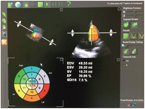
Fig. 1 Measurements of 3-dimensional left ventricular volumes using 3000 element matrix probe (3V probe) and analyzing the images of the ventricle using Tom Tec 4D Cardio-View RT 1.2 software. EDV: end-diastolic volume, ESV: end-systolic volume, SV: systolic volume, EF: ejection fraction, SDI: standard deviation index.
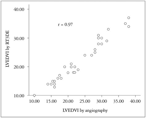
Fig. 2 Good correlation between real time three dimensional echocardiography (RT3DE) and cineangiography for estimation of left ventricular end diastolic volume index (LVEDVI) in all studied patients.

Fig. 3 Bland-Altman plot showing comparison between the left ventricular end diastolic volume index (LVEDVI) by real time three dimensional echocardiography and angiography.
Reference
-
1. Riehle TJ, Mahle WT, Parks WJ, Sallee D 3rd, Fyfe DA. Real-time three-dimensional echocardiographic acquisition and quantification of left ventricular indices in children and young adults with congenital heart disease: comparison with magnetic resonance imaging. J Am Soc Echocardiogr. 2008; 21:78–83.2. Monaghan MJ. Role of real time 3D echocardiography in evaluating the left ventricle. Heart. 2006; 92:131–136.3. Sugeng L, Weinert L, Lang RM. Left ventricular assessment using real time three dimensional echocardiography. Heart. 2003; 89:Suppl 3. iii29–iii36.4. Bu L, Munns S, Zhang H, Disterhoft M, Dixon M, Stolpen A, Sonka M, Scholz TD, Mahoney LT, Ge S. Rapid full volume data acquisition by real-time 3-dimensional echocardiography for assessment of left ventricular indexes in children: a validation study compared with magnetic resonance imaging. J Am Soc Echocardiogr. 2005; 18:299–305.5. Mannaerts HF, Van Der Heide JA, Kamp O, Papavassiliu T, Marcus JT, Beek A, Van Rossum AC, Twisk J, Visser CA. Quantification of left ventricular volumes and ejection fraction using freehand transthoracic three-dimensional echocardiography: comparison with magnetic resonance imaging. J Am Soc Echocardiogr. 2003; 16:101–109.6. Jenkins C, Bricknell K, Hanekom L, Marwick TH. Reproducibility and accuracy of echocardiographic measurements of left ventricular parameters using real-time three-dimensional echocardiography. J Am Coll Cardiol. 2004; 44:878–886.7. Sugeng L, Mor-Avi V, Weinert L, Niel J, Ebner C, Steringer-Mascherbauer R, Schmidt F, Galuschky C, Schummers G, Lang RM, Nesser HJ. Quantitative assessment of left ventricular size and function: side-by-side comparison of real-time three-dimensional echocardiography and computed tomography with magnetic resonance reference. Circulation. 2006; 114:654–661.8. Soriano BD, Hoch M, Ithuralde A, Geva T, Powell AJ, Kussman BD, Graham DA, Tworetzky W, Marx GR. Matrix-array 3-dimensional echocardiographic assessment of volumes, mass, and ejection fraction in young pediatric patients with a functional single ventricle: a comparison study with cardiac magnetic resonance. Circulation. 2008; 117:1842–1848.9. Iino M, Shiraishi H, Ichihashi K, Hoshina M, Saitoh M, Hirakubo Y, Morimoto Y, Momoi MY. Volume measurement of the left ventricle in children using real-time three-dimensional echocardiography: comparison with ventriculography. J Cardiol. 2007; 49:221–229.10. Greene DG, Carlisle R, Grant C, Bunnell IL. Estimation of left ventricular volume by one-plane cineangiography. Circulation. 1967; 35:61–69.11. Graham TP Jr, Faulkner S, Bender H Jr, Wender CM. Hypoplasia of the left ventricle: rare cause of postoperative mortality in tetralogy of Fallot. Am J Cardiol. 1977; 40:454–457.12. Matsuda H, Hirose H, Nakano S, Kishimoto H, Kato H, Kobayashi J, Miura T, Kato M, Kawashima Y. Age-related changes in right and left ventricular function in tetralogy of Fallot. Jpn Circ J. 1986; 50:1040–1043.13. Fukuda J, Izumi T, Matsukawa T, Eguchi S. Development of left ventricular muscle in tetralogy of Fallot. Jpn Circ J. 1984; 48:465–473.14. Heusch A, Rübo J, Krogmann ON, Bönig H, Bourgeois M. Volume measurement of the left ventricle in children with congenital heart defects: 3-dimensional echocardiography versus angiocardiography. Cardiology. 1999; 92:45–52.15. Kaye HH, Tynan M, Hunter S. Validity of echocardiographic estimates of left ventricular size and performance in infants and children. Br Heart J. 1975; 37:371–375.16. Lang RM, Badano LP, Tsang W, Adams DH, Agricola E, Buck T, Faletra FF, Franke A, Hung J, de Isla LP, Kamp O, Kasprzak JD, Lancellotti P, Marwick TH, McCulloch ML, Monaghan MJ, Nihoyannopoulos P, Pandian NG, Pellikka PA, Pepi M, Roberson DA, Shernan SK, Shirali GS, Sugeng L, Ten Cate FJ, Vannan MA, Zamorano JL, Zoghbi WA. American Society of Echocardiography. European Association of Echocardiography. EAE/ASE recommendations for image acquisition and display using three-dimensional echocardiography. Eur Heart J Cardiovasc Imaging. 2012; 13:1–46.
- Full Text Links
-
- Actions
-
Cited
- CITED
-
- Close
- Share
-
- Similar articles
-
- Echocardiographic Findings in Tetralogy of Fallot
- Volumetric Quantitation of Pulmonary Regurgitation and Right Ventricular Function in Postoperative Tetralogy of Fallot by Echocardiography and Magnetic Resonance Imaging
- Angiocardiographic diagnosis in tetralogy of Fallot
- Exercise tolerance tests in patients with tetralogy of Fallot repaired earlier: correlation with 2-dimensional echocardiography and cardiac catheterization
- Morphometric Analysis of the Infundibulum in Tetralogy of Fallot
