Influence of Heart Rate and Innovative Motion-Correction Algorithm on Coronary Artery Image Quality and Measurement Accuracy Using 256-Detector Row Computed Tomography Scanner: Phantom Study
- Affiliations
-
- 1Department of Radiology, Pusan National University School of Medicine and Medical Research Institute, Pusan National University Hospital, Busan, Korea. jw@pusan.ac.kr
- KMID: 2429923
- DOI: http://doi.org/10.3348/kjr.2018.0251
Abstract
OBJECTIVE
To investigate the efficacy of motion-correction algorithm (MCA) in improving coronary artery image quality and measurement accuracy using an anthropomorphic dynamic heart phantom and 256-detector row computed tomography (CT) scanner.
MATERIALS AND METHODS
An anthropomorphic dynamic heart phantom was scanned under a static condition and under heart rate (HR) simulation of 50-120 beats per minute (bpm), and the obtained images were reconstructed using conventional algorithm (CA) and MCA. We compared the subjective image quality of coronary arteries using a four-point scale (1, excellent; 2, good; 3, fair; 4, poor) and measurement accuracy using measurement errors of the minimal luminal diameter (MLD) and minimal luminal area (MLA).
RESULTS
Compared with CA, MCA significantly improved the subjective image quality at HRs of 110 bpm (1.3 ± 0.3 vs. 1.9 ± 0.8, p = 0.003) and 120 bpm (1.7 ± 0.7 vs. 2.3 ± 0.6, p = 0.006). The measurement error of MLD significantly decreased on using MCA at 110 bpm (11.7 ± 5.9% vs. 18.4 ± 9.4%, p = 0.013) and 120 bpm (10.0 ± 7.3% vs. 25.0 ± 16.5%, p = 0.013). The measurement error of the MLA was also reduced using MCA at 110 bpm (19.2 ± 28.1% vs. 26.4 ± 21.6%, p = 0.028) and 120 bpm (17.9 ± 17.7% vs. 34.8 ± 19.6%, p = 0.018).
CONCLUSION
Motion-correction algorithm can improve the coronary artery image quality and measurement accuracy at a high HR using an anthropomorphic dynamic heart phantom and 256-detector row CT scanner.
Keyword
Figure
-
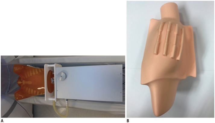
Fig. 1 Anthropomorphic dynamic heart phantom and simulated heart with stenotic model.A. Anthropomorphic dynamic heart phantom. B. Simulated heart with stenotic model.
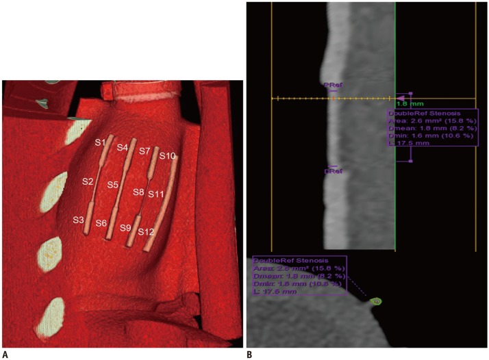
Fig. 2 Volume-rendered curved multiplanar reformatted CT images and transverse section of coronary artery demonstrating segmentations of coronary arteries and measurement of MLD and MLA.A. Volume-rendering images of heart phantom at HR of 70 bpm demonstrate segmentations of coronary artery. There are 4 coronary arteries in cardiac phantom. Each coronary artery was divided into three segments. Therefore, total of 12 coronary artery segments were analyzed. B. Curved multiplanar reformatted CT images and transverse section of S2 illustrating method used to measure MLD and MLA. When PRef and DRef were set for each segment, software automatically measure MDL and MLA of each segment (yellow line represents narrowest level of S2). In case software presented incorrect centerline or outline, observer was able to adjust it. At HR of 70 bpm, MLD and MLA of S2 were 1.6 mm and 2.6 mm2, respectively. Data in parenthesis (%) represents stenosis percentage using dual reference selection. bpm = beats per minute, CT = computed tomography, Dmean = mean luminal diameter, Dmin = minimal luminal diameter, DRef = distal references, HR = heart rate, L = length, MLA = minimal luminal area, MLD = minimal luminal diameter, PRef = proximal references, S = segment
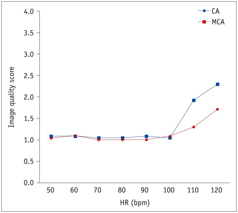
Fig. 3 Mean image quality scores of CA and MCA according to HR.Image quality score at HR of 120 bpm was significantly higher (all p values < 0.001) than those at 50–100 bpm according to Kruskal-Wallis test with following pair-wise comparison using Mann-Whitney test and Bonferroni correction. CA = conventional algorithm, MCA = motion correction algorithm
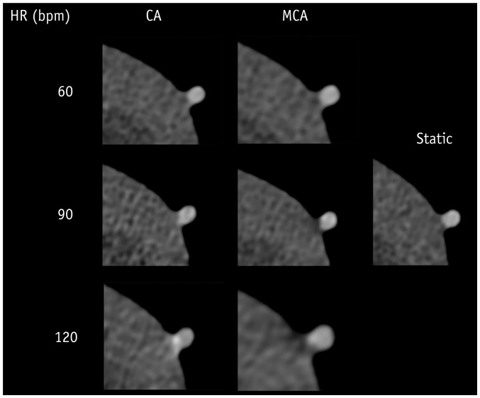
Fig. 4 Representative axial images reconstructed by CA and MCA at HRs of static and 60, 90, and 120 bpm.
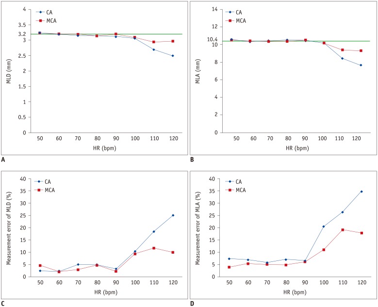
Fig. 5 Measurement accuracy according to HR between CA and MCA.A. MLD. Green line presents MLD at static (reference, 3.2 mm). B. MLA. Green line presents MLA at static (reference, 10.4 mm2). C. Measurement error of MLD. D. Measurement error of MLA.
Cited by 1 articles
-
Clinical Applications of Wide-Detector CT Scanners for Cardiothoracic Imaging: An Update
Eun-Ju Kang
Korean J Radiol. 2019;20(12):1583-1596. doi: 10.3348/kjr.2019.0327.
Reference
-
1. Schroeder S, Kopp AF, Kuettner A, Burgstahler C, Herdeg C, Heuschmid M, et al. Influence of heart rate on vessel visibility in noninvasive coronary angiography using new multislice computed tomography: experience in 94 patients. Clin Imaging. 2002; 26:106–111. PMID: 11852217.2. Hoffmann MH, Shi H, Manzke R, Schmid FT, De Vries L, Grass M, et al. Noninvasive coronary angiography with 16-detector row CT: effect of heart rate. Radiology. 2005; 234:86–97. PMID: 15550373.
Article3. Min JK, Arsanjani R, Kurabayashi S, Andreini D, Pontone G, Choi BW, et al. Rationale and design of the ViCTORY (Validation of an Intracycle CT Motion CORrection Algorithm for Diagnostic AccuracY) trial. J Cardiovasc Comput Tomogr. 2013; 7:200–206. PMID: 23849493.
Article4. Leipsic J, Labounty TM, Hague CJ, Mancini GB, O'Brien JM, Wood DA, et al. Effect of a novel vendor-specific motion-correction algorithm on image quality and diagnostic accuracy in persons undergoing coronary CT angiography without rate-control medications. J Cardiovasc Comput Tomogr. 2012; 6:164–171. PMID: 22551593.
Article5. Andreini D, Pontone G, Mushtaq S, Bertella E, Conte E, Segurini C, et al. Low-dose CT coronary angiography with a novel IntraCycle motion-correction algorithm in patients with high heart rate or heart rate variability. Eur Heart J Cardiovasc Imaging. 2015; 16:1093–1100. PMID: 25762564.
Article6. Liang J, Wang H, Xu L, Dong L, Fan Z, Wang R, et al. Impact of SSF on diagnostic performance of coronary computed tomography angiography within 1 heart beat in patients with high heart rate using a 256-row detector computed tomography. J Comput Assist Tomogr. 2018; 42:54–61. PMID: 28708724.
Article7. Farshad-Amacker NA, Alkadhi H, Leschka S, Frauenfelder T. Effect of high-pitch dual-source CT to compensate motion artifacts: a phantom study. Acad Radiol. 2013; 20:1234–1239. PMID: 24029055.8. Cho I, Elmore K, Ó Hartaigh B, Schulman-Marcus J, Granser H, Valenti V, et al. Heart-rate dependent improvement in image quality and diagnostic accuracy of coronary computed tomographic angiography by novel intracycle motion correction algorithm. Clin Imaging. 2015; 39:421–426. PMID: 25649255.
Article9. Toepker M, Euller G, Unger E, Weber M, Kienzl D, Herold CJ, et al. Stenosis quantification of coronary arteries in coronary vessel phantoms with second-generation dual-source CT: influence of measurement parameters and limitations. AJR Am J Roentgenol. 2013; 201:W227–W234. PMID: 23883237.
Article10. Wang YT, Yang CY, Hsiao JK, Liu HM, Lee WJ, Shen Y. The influence of reconstruction algorithm and heart rate on coronary artery image quality and stenosis detection at 64-detector cardiac CT. Korean J Radiol. 2009; 10:227–234. PMID: 19412510.
Article11. ICRP. The 2007 recommendations of the International Commission on Radiological Protection. ICRP Publication 103. Ann ICRP. 2007; 37:1–332.12. Koo TK, Li MY. A guideline of selecting and reporting intraclass correlation coefficients for reliability research. J Chiropr Med. 2016; 15:155–163. PMID: 27330520.
Article13. Abbara S, Blanke P, Maroules CD, Cheezum M, Choi AD, Han BK, et al. SCCT guidelines for the performance and acquisition of coronary computed tomographic angiography: a report of the Society of Cardiovascular Computed Tomography Guidelines Committee: endorsed by the North American Society for Cardiovascular Imaging (NASCI). J Cardiovasc Comput Tomogr. 2016; 10:435–449. PMID: 27780758.14. Achenbach S, Marwan M, Schepis T, Pflederer T, Bruder H, Allmendinger T, et al. High-pitch spiral acquisition: a new scan mode for coronary CT angiography. J Cardiovasc Comput Tomogr. 2009; 3:117–121. PMID: 19332343.
Article15. Rybicki FJ, Otero HJ, Steigner ML, Vorobiof G, Nallamshetty L, Mitsouras D, et al. Initial evaluation of coronary images from 320-detector row computed tomography. Int J Cardiovasc Imaging. 2008; 24:535–546. PMID: 18368512.
Article16. Achenbach S, Ropers U, Kuettner A, Anders K, Pflederer T, Komatsu S, et al. Randomized comparison of 64-slice single- and dual-source computed tomography coronary angiography for the detection of coronary artery disease. JACC Cardiovasc Imaging. 2008; 1:177–186. PMID: 19356426.
Article17. Xing Y, Zhao Y, Guo N, Pan CX, Azati G, Wang YW, et al. Effect of a novel intracycle motion correction algorithm on dual-energy spectral coronary CT angiography: a study with pulsating coronary artery phantom at high heart rates. Korean J Radiol. 2017; 18:881–887. PMID: 29089820.
Article18. Li ZN, Yin WH, Lu B, Yan HB, Mu CW, Gao Y, et al. Improvement of image quality and diagnostic performance by an innovative motion-correction algorithm for prospectively ECG triggered coronary CT angiography. PLoS One. 2015; 10:e0142796. PMID: 26571417.
Article19. Lee H, Kim JA, Lee JS, Suh J, Paik SH, Park JS. Impact of a vendor-specific motion-correction algorithm on image quality, interpretability, and diagnostic performance of daily routine coronary CT angiography: influence of heart rate on the effect of motion-correction. Int J Cardiovasc Imaging. 2014; 30:1603–1612. PMID: 25038955.
Article
- Full Text Links
-
- Actions
-
Cited
- CITED
-
- Close
- Share
-
- Similar articles
-
- Effect of a Novel Intracycle Motion Correction Algorithm on Dual-Energy Spectral Coronary CT Angiography: A Study with Pulsating Coronary Artery Phantom at High Heart Rates
- The Influence of Reconstruction Algorithm and Heart Rate on Coronary Artery Image Quality and Stenosis Detection at 64-Detector Cardiac CT
- 16-Slice Multi-Detector Row CT Coronary Angiography: Image Quality and Optimization of the Image Reconstruction Window
- Coronary CT Angiography
- Unusual Coronary Artery Fistula: Left Anterior Descending Coronary Artery - Left Ventricular Fistula Diagnosed by ECG-Gated Multi-Detector Row Coronary CT Angiography
