Ann Dermatol.
2016 Feb;28(1):138-139. 10.5021/ad.2016.28.1.138.
Secondary Granulomatous Cutaneous Involvement in Peripheral T-cell Lymphoma
- Affiliations
-
- 1Department of Dermatology, Yeungnam University College of Medicine, Daegu, Korea. dhshin@med.yu.ac.kr
- KMID: 2429504
- DOI: http://doi.org/10.5021/ad.2016.28.1.138
Abstract
- No abstract available.
MeSH Terms
Figure
-
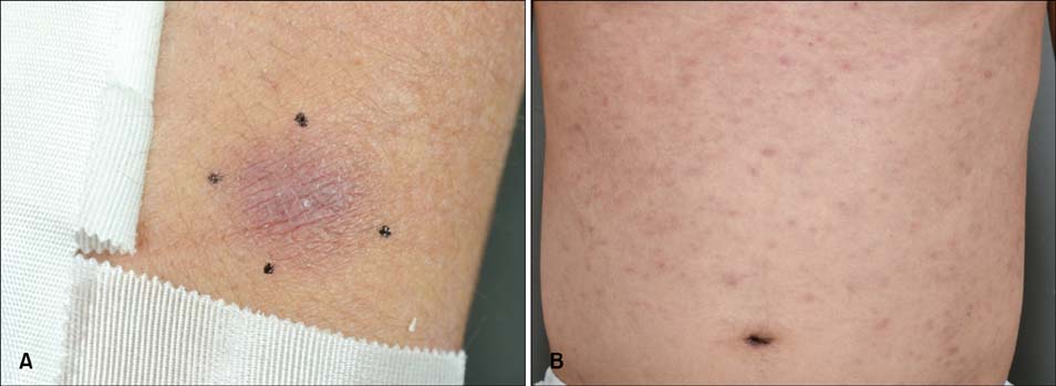
Fig. 1 (A) A 2×2-cm-sized hyperpigmented nodule on the left arm. (B) Multiple miliary grain to rice-grain-sized erythematous to brownish macules and papules on the trunk.
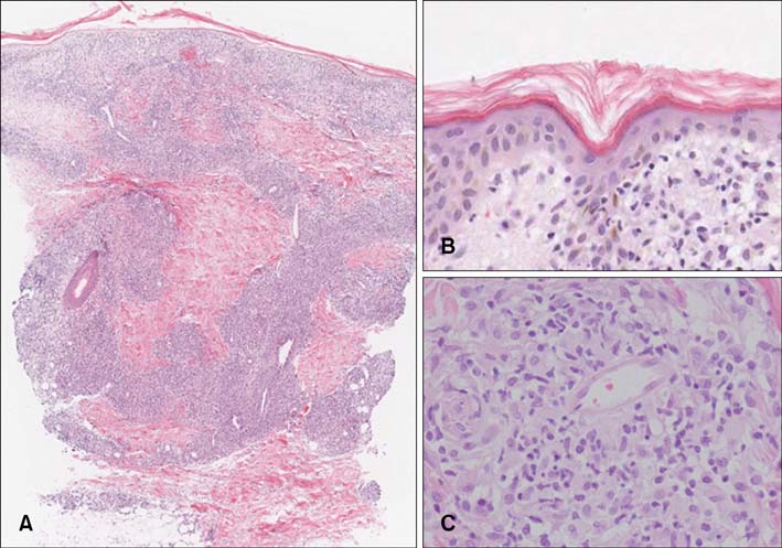
Fig. 2 (A) Dermal, perivascular nodular inflammatory cell infiltration. (B) Epidermotropism of lymphocytes, with hyperchromatic and indented nucleus. (C) Many histiocytes and atypical lymphocytes with hyperchromatic nuclei form sarcoid-like granuloma (H&E; A: ×40, B: ×200, C: ×200).
Reference
-
1. Gallardo F, García-Muret MP, Servitje O, Estrach T, Bielsa I, Salar A, et al. Cutaneous lymphomas showing prominent granulomatous component: clinicopathological features in a series of 16 cases. J Eur Acad Dermatol Venereol. 2009; 23:639–647.
Article2. Pujol RM, Gallardo F, Servitje O, Martí RM, Bordes R, García-Muret MP, et al. Peripheral T-cell lymphoma with secondary epithelioid granulomatous cutaneous involvement: a clinicopathologic study of four cases. J Dermatol. 2005; 32:541–548.
Article3. Patsouris E, Noël H, Lennert K. Histological and immunohistological findings in lymphoepithelioid cell lymphoma (Lennert's lymphoma). Am J Surg Pathol. 1988; 12:341–350.
Article4. Flaxman BA, Koumans JA, Ackerman AB. Granulomatous mycosis fungoides. A 14-year follow-up of a case. Am J Dermatopathol. 1983; 5:145–151.
- Full Text Links
-
- Actions
-
Cited
- CITED
-
- Close
- Share
-
- Similar articles
-
- A Case of Primary Nasal CD56+ NK/T cell Lymphoma with Cutaneous Involvement
- Case of Secondary Cutaneous Peripheral T Cell Lymphoma, NOS
- A Case of Secondary Cutaneous Diffuse Large B-cell Lymphoma
- A Case of Cutaneous T Cell Lymphoma Presenting as Papuloerythroderma of Ofuji
- A Case of Aggressive T/NK-cell Lymphoma/leukemia with Cutaneous Involvement
