Blood Res.
2018 Dec;53(4):281-287. 10.5045/br.2018.53.4.281.
Detection of bone marrow involvement with FDG PET/CT in patients with newly diagnosed lymphoma
- Affiliations
-
- 1Bloodworks Research Institute, Seattle, WA, USA. tahsino@bloodworksnw.org
- 2Department of Nuclear Medicine, Istanbul School of Medicine, Istanbul, Turkey.
- 3Department of Hematology, Istanbul School of Medicine, Istanbul, Turkey.
- 4Department of Biostatistics and Epidemiology, Marmara School of Medicine, Istanbul, Turkey.
- 5Department of Pathology, Istanbul School of Medicine, Istanbul, Turkey.
- KMID: 2429301
- DOI: http://doi.org/10.5045/br.2018.53.4.281
Abstract
- BACKGROUND
Bone marrow involvement (BMI) affects the lymphoma stage, survival, and treatment. Bone marrow biopsy (BMB) and fluorodeoxyglucose (FDG) positron emission tomography- computed tomography (PET/CT) are useful techniques to detect BMI. Both have advantages and disadvantages. We aimed to identify factors that could be used to predict BMI with positive and negative results on PET/CT compare them with BMB in newly diagnosed patients with lymphoma.
METHODS
We included 22 non-Hodgkin and 16 Hodgkin lymphoma patients in this single center study. All patients had PET/CT examination and BMB before treatment. BMI in BMB was reported as negative or positive. Bone marrow was classified into 3 types by FDG uptake on PT/CT; diffuse involvement, focal involvement, and normal bone marrow.
RESULTS
PET/CT and BMB results were concordant (7 positive, 15 negative) in 22 patients (57%). We evaluated concordant and discordant patient characteristics and risk-stratified patients for BMI. Our findings suggest that patients with diffuse FDG uptake on PET/CT, especially patients with advanced age and low platelet and white blood cell counts, are likely to have BMI and could potentially forego BMB. Patients with negative PET/CT findings and no significant laboratory abnormalities are very unlikely to have BMI.
CONCLUSION
Our results suggest that BMI should not be decided solely based PET/CT or BMB findings. It is reasonable to use both diagnostic assays along with clinical and laboratory findings. PET/CT result, clinical and laboratory findings could be useful for predicting BMI in patient for whom BMB is contraindicated.
MeSH Terms
Figure
-
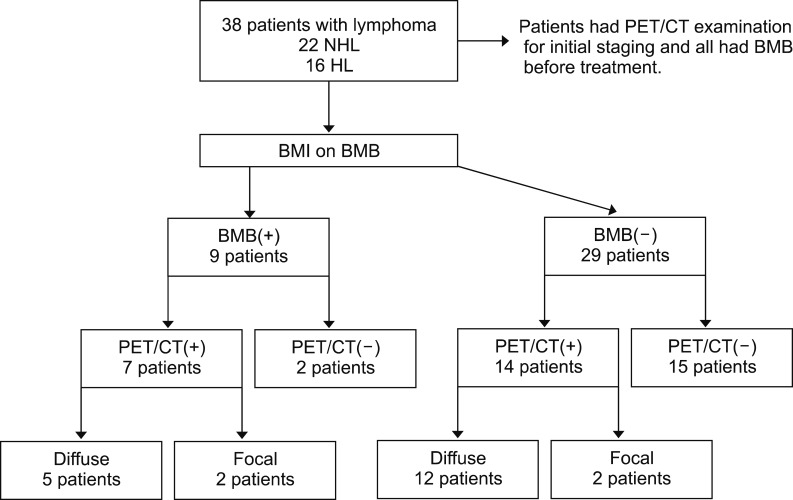
Fig. 1 The study flow chart.Abbreviations: BMB, bone marrow biopsy; BMI, bone marrow involvement; HL, Hodgkin's lymphoma; NHL, non-Hodgkin lymphoma; PET/CT, Positron emission tomography-computed tomography.
Reference
-
1. Moog F, Bangerter M, Diederichs CG, et al. Extranodal malignant lymphoma: detection with FDG PET versus CT. Radiology. 1998; 206:475–481. PMID: 9457202.
Article2. Even-Sapir E. Imaging of malignant bone involvement by morphologic, scintigraphic, and hybrid modalities. J Nucl Med. 2005; 46:1356–1367. PMID: 16085595.3. El Karak F, Bou-Orm IR, Ghosn M, et al. PET/CT scanner and bone marrow biopsy in detection of bone marrow involvement in diffuse large B-cell lymphoma. PLoS One. 2017; 12:e0170299. PMID: 28099514.
Article4. Solal-Céligny P, Roy P, Colombat P, et al. Follicular lymphoma international prognostic index. Blood. 2004; 104:1258–1265. PMID: 15126323.
Article5. Hasenclever D, Diehl V. A prognostic score for advanced Hodgkin's disease. International Prognostic Factors Project on Advanced Hodgkin's Disease. N Engl J Med. 1998; 339:1506–1514. PMID: 9819449.6. Federico M, Bellei M, Marcheselli L, et al. Follicular lymphoma international prognostic index 2: a new prognostic index for follicular lymphoma developed by the international follicular lymphoma prognostic factor project. J Clin Oncol. 2009; 27:4555–4562. PMID: 19652063.
Article7. National Cancer Institute. Survival. Bethesda, MD: National Cancer Institute;2018. Accessed October 2, 2018. at https://progressreport.cancer.gov/after/survival.8. Pui CH, Thiel E. Central nervous system disease in hematologic malignancies: historical perspective and practical applications. Semin Oncol. 2009; 36(4 Suppl 2):S2–S16. PMID: 19660680.
Article9. Fueger BJ, Yeom K, Czernin J, Sayre JW, Phelps ME, Allen-Auerbach MS. Comparison of CT, PET, and PET/CT for staging of patients with indolent non-Hodgkin's lymphoma. Mol Imaging Biol. 2009; 11:269–274. PMID: 19326177.
Article10. Fuertes S, Setoain X, López-Guillermo A, et al. The value of positron emission tomography/computed tomography (PET/CT) in the staging of diffuse large B-cell lymphoma. Med Clin (Barc). 2007; 129:688–693. PMID: 18021609.11. Freudenberg LS, Antoch G, Schütt P, et al. FDG-PET/CT in re-staging of patients with lymphoma. Eur J Nucl Med Mol Imaging. 2004; 31:325–329. PMID: 14647988.
Article12. McKenna RW, Bloomfield CD, Brunning RD. Nodular lymphoma: bone marrow and blood manifestations. Cancer. 1975; 36:428–440. PMID: 50871.
Article13. Mckenna RW. The bone marrow manifestations of Hodgkin's disease, the non-Hodgkin's lymphomas and lymphoma-like disorders. In : Dunphy CH, editor. Neoplastic hematopathology. 1st ed. Baltimore, MD: Williams & Wilkins;1992. p. 1156.14. Elstrom RL, Tsai DE, Vergilio JA, Downs LH, Alavi A, Schuster SJ. Enhanced marrow [18F]fluorodeoxyglucose uptake related to myeloid hyperplasia in Hodgkin's lymphoma can simulate lymphoma involvement in marrow. Clin Lymphoma. 2004; 5:62–64. PMID: 15245610.
Article15. Aflalo-Hazan V, Gutman F, Kerrou K, Montravers F, Grahek D, Talbot JN. Increased FDG uptake by bone marrow in major beta-thalassemia. Clin Nucl Med. 2005; 30:754–755. PMID: 16237306.
Article16. Murata Y, Kubota K, Yukihiro M, Ito K, Watanabe H, Shibuya H. Correlations between 18F-FDG uptake by bone marrow and hematological parameters: measurements by PET/CT. Nucl Med Biol. 2006; 33:999–1004. PMID: 17127173.
Article17. El-Galaly TC, d'Amore F, Mylam KJ, et al. Routine bone marrow biopsy has little or no therapeutic consequence for positron emission tomography/computed tomography-staged treatment-naive patients with Hodgkin lymphoma. J Clin Oncol. 2012; 30:4508–4514. PMID: 23150698.
Article18. Adams HJ, Kwee TC, Fijnheer R, Dubois SV, Nievelstein RA, de Klerk JM. Diffusely increased bone marrow FDG uptake in recently untreated lymphoma: incidence and relevance. Eur J Haematol. 2015; 95:83–89. PMID: 25537478.
Article19. Pakos EE, Fotopoulos AD, Ioannidis JP. 18F-FDG PET for evaluation of bone marrow infiltration in staging of lymphoma: a meta-analysis. J Nucl Med. 2005; 46:958–963. PMID: 15937306.20. Buchmann I, Reinhardt M, Elsner K, et al. 2-(fluorine-18) fluoro-2-deoxy-D-glucose positron emission tomography in the detection and staging of malignant lymphoma. A bicenter trial. Cancer. 2001; 91:889–899. PMID: 11251940.21. Carr R, Barrington SF, Madan B, et al. Detection of lymphoma in bone marrow by whole-body positron emission tomography. Blood. 1998; 91:3340–3346. PMID: 9558391.
Article
- Full Text Links
-
- Actions
-
Cited
- CITED
-
- Close
- Share
-
- Similar articles
-
- Usefulness of 18F-FDG PET/CT for the Evaluation of Bone Marrow Involvement in Patients with High-Grade Non-Hodgkin's Lymphoma
- The Clinical Utility of FDG PET-CT in Evaluation of Bone Marrow Involvement by Lymphoma
- Detection of Lymphomatous Marrow Infiltration using F-18 FDG PET at Initial Staging and after chemotherapy
- Prognostic Value of Bone Marrow F-18 FDG Uptake in Patients with Advanced-Stage Diffuse Large B-Cell Lymphoma
- 18F-Fluorodeoxyglucose-Positron Emission Tomography in the Staging of Malignant Lymphoma Compared with CT and 67Ga Scan
