Clin Exp Otorhinolaryngol.
2018 Sep;11(3):174-180. 10.21053/ceo.2017.00864.
Evaluation of the Possible Neurotoxic Effect of the Bone Cement on the Facial Nerve: An Experimental Study
- Affiliations
-
- 1Department of Otorhinolaryngology, Goztepe Training and Research Hospital, Istanbul Medeniyet University, Faculty of Medicine, Istanbul, Turkey. mtkalcioglu@hotmail.com
- 2Department of Otorhinolaryngology, Sakarya University Training and Research Hospital, Sakarya, Turkey.
- 3Department of Physiology, Istanbul Medeniyet University, Faculty of Medicine, Istanbul, Turkey.
- 4Department of Pathology, Yeditepe University, Faculty of Medicine, Istanbul, Turkey.
- KMID: 2429006
- DOI: http://doi.org/10.21053/ceo.2017.00864
Abstract
OBJECTIVES
To investigate neurotoxic effect of bone cement (BC) on facial nerve by using electrophysiological and histopathological methods.
METHODS
This study included 20 male albino Wistar rats, divided into four equal groups. Group A was designed as the control group, while group B was sham group. In the group C, BC solution was dropped onto the facial nerve trunks of rats and washed with physiological saline after 5 seconds. In the group D, BC solution was dropped onto the facial nerve trunks of rats and after allowing 5 minutes to dry, wounds were closed. Pre- and postoperative (on 4th week) evoked electromyography (EMG) measurements were done. For histopathological assessments, the rats were euthanized and tissue samples of facial nerve and surrounding areas were collected.
RESULTS
According to the wave amplitude levels of evoked EMG, postoperative amplitude levels of group D were significantly decreased, compared to preoperative amplitude levels (P=0.043). We found no statistically significant difference in inflammation among the groups. In none of the groups, foreign body reaction and granulation tissue were not detected in any of the groups. In addition, degeneration in axon, myelin, or perineural nets was not detected in any of the groups.
CONCLUSION
This study results suggest that BC has no direct toxicity on facial nerve, while it has indirect effects, by decreasing amplitude. Therefore, we conclude that direct contact of BC with nerve should be avoided, and the area should be cleaned by aspiration or washing with physiological saline in case of contact.
Keyword
MeSH Terms
Figure
-
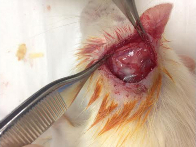
Fig. 1. Group D: bone cement was dropped and kept for 5 minutes drying.

Fig. 2. Histopathologic section of the facial nerves and surrounding tissues of rats in all groups: H&E and s-100 stain (left and right, respectively), scale bar indicates 100 μm. The section is from (A) group A (control), (B) group B (sham), (C) group C (bone cement [BC] is applied for 5 seconds and cleaned), and (D) from group D (BC was applied and kept for 5 minutes drying). There was no axonal degeneration in all figures. Perineural sheath and myelin structure layout were also regular (shown with arrows), but there was an inflammation of muscle and myositis in a rat tissue of group D (shown with asterisk).
Reference
-
1. Righini-Grunder F, Hausler R, Chongvisal S, Caversaccio M. Glass ionomer cement in otological microsurgery: experience over 16 years. Eur Arch Otorhinolaryngol. 2015; Oct. 272(10):2749–54.2. Hafiz G. A more reliable method for incudostapedial rebridging ossiculoplasty: bone cement and wire. Adv Ther. 2005; Jan-Feb. 22(1):56–62.
Article3. Kalcioglu MT, Tan M, Fleerakkers J. The use of bone cement for ossicular chain defects. Eur Arch Otorhinolaryngol. 2013; Nov. 270(11):2849–55.
Article4. Kalcioglu MT, Uzun IH, Yalcin M, Malkoc MA, Ogreten AT, Hanege FM. Evaluation on shear bond strength of different glass ionomer and hydroxy apatite cements used in ossiculoplasty. Balkan Med J. 2015; Jan. 32(1):23–9.
Article5. Guillard O, Pineau A, Fauconneau B, Chobaut JC, Desaulty A, Angot A, et al. Biological levels of aluminium after use of aluminium-containing bone cement in post-otoneurosurgery. J Trace Elem Med Biol. 1997; Apr. 11(1):53–6.
Article6. Kanjevac TV, Milovanovic MZ, Milosevic-Djordjevic O, Tesic Z, Ivanovic M, Lukic A. Cytotoxicity of glass ionomer cement on human exfoliated deciduous teeth stem cells correlates with released fluoride, strontium and aluminum ion concentrations. Arch Biol Sci. 2015; 67(2):619–30.
Article7. Granstrom G, Holmquist J, Tjellstrom A. Facial nerve paralysis following repair of the external ear canal with ionomeric cement. Ear Nose Throat J. 2000; Jul. 79(7):495–8.
Article8. Renard JL, Felten D, Bequet D. Post-otoneurosurgery aluminium encephalopathy. Lancet. 1994; Jul. 344(8914):63–4.
Article9. Reusche E, Pilz P, Oberascher G, Lindner B, Egensperger R, Gloeckner K, et al. Subacute fatal aluminum encephalopathy after reconstructive otoneurosurgery: a case report. Hum Pathol. 2001; Oct. 32(10):1136–40.
Article10. Brook IM, Hatton PV. Glass-ionomers: bioactive implant materials. Biomaterials. 1998; Mar. 19(6):565–71.
Article11. Hatton PV, Hurrell-Gillingham K, Brook IM. Biocompatibility of glass-ionomer bone cements. J Dent. 2006; Sep. 34(8):598–601.
Article12. Murai N, Oda Y, Nagata I, Takahashi JA, Ishikawa M, Kikuchi H, et al. Neurotoxicity testing of a new bioactive bone cement. Neurol Med Chir (Tokyo). 1997; Feb. 37(2):201–4.
Article13. Kosar AT. Neurotoxic effect of glass ionomer cement: experimental study. Medical speciality thesis [Internet]. Istanbul: The Ministry of Health of Turkey, Sisli Etfal Training and Research Hospital, Department of Otorhinolaryngology;2008 [cited 2018 Mar 1]. Available from: http://www.istanbulsaglik.gov.tr/w/tez/pdf/kbb/dr_alper_tolga_kosar.pdf.14. Salomone R, Costa HJ, Rodrigues JR, Reis e Silva SM, Ovando PC, Bento RF. Assessment of a neurophysiological model of the mandibular branch of the facial nerve in rats by electromyography. Ann Otol Rhinol Laryngol. 2012; Mar. 121(3):179–84.
Article15. Nicholson JW, Czarnecka B. Review paper: role of aluminum in glass-ionomer dental cements and its biological effects. J Biomater Appl. 2009; Nov. 24(4):293–308.
Article16. Aranha AM, Giro EM, Souza PP, Hebling J, de Souza Costa CA. Effect of curing regime on the cytotoxicity of resin-modified glass-ionomer lining cements applied to an odontoblast-cell line. Dent Mater. 2006; Sep. 22(9):864–9.
Article17. Ribeiro DA, Marques ME, Salvadori DM. Biocompatibility of glassionomer cements using mouse lymphoma cells in vitro. J Oral Rehabil. 2006; Dec. 33(12):912–7.
Article18. Meryon SD, Stephens PG, Browne RM. A comparison of the in vitro cytotoxicity of two glass-ionomer cements. J Dent Res. 1983; Jun. 62(6):769–73.
Article19. Kong N, Jiang T, Zhou Z, Fu J. Cytotoxicity of polymerized resin cements on human dental pulp cells in vitro. Dent Mater. 2009; Nov. 25(11):1371–5.
Article20. Souza PP, Aranha AM, Hebling J, Giro EM, Costa CA. In vitro cytotoxicity and in vivo biocompatibility of contemporary resin-modified glass-ionomer cements. Dent Mater. 2006; Sep. 22(9):838–44.
Article21. Nguyen Ngoc TD, Son YO, Lim SS, Shi X, Kim JG, Heo JS, et al. Sodium fluoride induces apoptosis in mouse embryonic stem cells through ROS-dependent and caspase- and JNK-mediated pathways. Toxicol Appl Pharmacol. 2012; Mar. 259(3):329–37.
Article22. Ramsden RT, Herdman RC, Lye RH. Ionomeric bone cement in neuro-otological surgery. J Laryngol Otol. 1992; Nov. 106(11):949–53.
Article23. Kupperman D, Tange RA. Ionomeric cement in the human middle ear cavity: long-term results of 23 cases. Laryngoscope. 2001; Feb. 111(2):306–9.
Article24. Lee DH. Clinical efficacy of electroneurography in acute facial paralysis. J Audiol Otol. 2016; Apr. 20(1):8–12.
Article25. Bayazit YA, Ozer E, Kanlikama M, Durmaz T, Yilmaz M. Bone cement ossiculoplasty: incus to stapes versus malleus to stapes cement bridge. Otol Neurotol. 2005; May. 26(3):364–7.
Article26. Babighian G. Use of a glass ionomer cement in otological surgery: a preliminary report. J Laryngol Otol. 1992; Nov. 106(11):954–9.
Article
- Full Text Links
-
- Actions
-
Cited
- CITED
-
- Close
- Share
-
- Similar articles
-
- An Experimental Study of the Effects of Cementing Stage and the Presence of Synovial Fluid and Physiologic Saline at the Interface upon the Tensile Bonding Strength of Acrylic Bone Cement
- Facial Nerve Paralysis and Surgical Management
- Report of facial nerve decompression in the traumatic facial palsy
- A Study on The Antibacterial Effect of antibiotic Impregnated Bone Cement
- Bilateral Bifurcation of the Mastoid Segment of the Facial Nerve
