Ann Dermatol.
2018 Dec;30(6):749-751. 10.5021/ad.2018.30.6.749.
Syphilitic Gumma: A Rare Form of Cutaneous Tertiary Syphilis
- Affiliations
-
- 1Department of Dermatology, SMG-SNU Boramae Medical Center, Seoul, Korea. snuhdm@gmail.com
- KMID: 2428943
- DOI: http://doi.org/10.5021/ad.2018.30.6.749
Abstract
- No abstract available.
MeSH Terms
Figure
-

Fig. 1 (A) A nontender dusky-red to brown plaque with deep, centrally punched-out ulcers covered with some crusts and necrotic debris. (B) A significant improvement shown after three intramuscular injections of penicillin G.
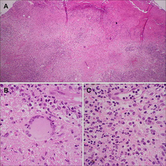
Fig. 2 (A) Extensive necrosis and granulomatous inflammation within the dermis (H&E, ×40), including dense infiltrates composed predominantly of (B) multinucleated giant cells (H&E, ×400), (C) lymphocytes and plasma cells (H&E, ×400).
Reference
-
1. Benzaquen M, Horreau C, Koeppel MC, Berbis P. A pseudotumoral facial mass revealing tertiary syphilis. Clin Exp Dermatol. 2017; 42:714–716.
Article2. Rocha N, Horta M, Sanches M, Lima O, Massa A. Syphilitic gumma--cutaneous tertiary syphilis. J Eur Acad Dermatol Venereol. 2004; 18:517–518.3. Boyd AS. Syphilitic gumma arising in association with foreign material. J Cutan Pathol. 2016; 43:1028–1030.
Article4. Masege SD, Karstaedt A. A rare case of a chronic syphilitic gumma in a man infected with human immunodeficiency virus. J Laryngol Otol. 2014; 128:557–560.
Article5. Leão JC, Gueiros LA, Porter SR. Oral Manifestations of Syphilis. Clinics (Sao Paulo). 2006; 61:161–166.
Article
- Full Text Links
-
- Actions
-
Cited
- CITED
-
- Close
- Share
-
- Similar articles
-
- Cerebral Gumma Mimicking a Brain Tumor in a Human Immunodeficiency Virus-Negative Patient: A Case Report
- Cerebral Syphilitic Gumma Mimicking a Brain Tumor in the Relapse of Secondary Syphilis in a Human Immunodeficiency Virus-Negative Patient
- Three Cases of Secondary Syphilis with Simultaneous Primary Syphilitic Lesions
- A Case of Syphilitic Keratoderma Concurrent with Syphilitic Uveitis
- 3 Cases of Interstitial Keratitis Occurred in Congenital Syphilitic Patients
