Perforating Granuloma Annulare Mimicking Papulonecrotic Tuberculid
- Affiliations
-
- 1Department of Dermatology, Medical Research Institute, Chungbuk National University School of Medicine, Cheongju, Korea. tyyoon@chungbuk.ac.kr
- KMID: 2428930
- DOI: http://doi.org/10.5021/ad.2018.30.6.716
Abstract
- Perforating granuloma annulare (PGA), a rare variant of granuloma annulare, is characterized by transepidermal elimination of altered collagen that clinically manifests an umbilicated papule with a central crust. It can be confused with papulonecrotic tuberculid (PNT) because of their similar appearance. Unlike PGA, PNT is usually related to tuberculosis infection with a typical histologic finding of wedge-shaped dermal necrosis. Here, we report the first Korean case of PGA mimicking PNT both clinically and histologically. A 43-year-old Korean woman presented with erythematous papules localized on the extensor surface of her limbs for one year. Some of these papules had a central umbilication or a crust. Regarding comorbidity, she had latent tuberculosis diagnosed with QuantiFERON®-TB Gold test about five months ago. She was on antituberculous medication. Initially, a diagnosis of papulonecrotic tuberculid accompanied by latent tuberculosis was considered. However, despite taking the antituberculous medication for five months, her skin lesions were not improved. Biopsy specimen from her arm lesion showed wedge-shaped area of necrosis in the dermis. Additionally, there were multiple focal mucin depositions and palisading granulomatous inflammation throughout the dermis. A diagnosis of PGA was made and she was treated with topical corticosteroid. After two weeks of applying topical corticosteroid, most of her skin lesions disappeared, leaving some hyperpigmented scars.
MeSH Terms
Figure
-
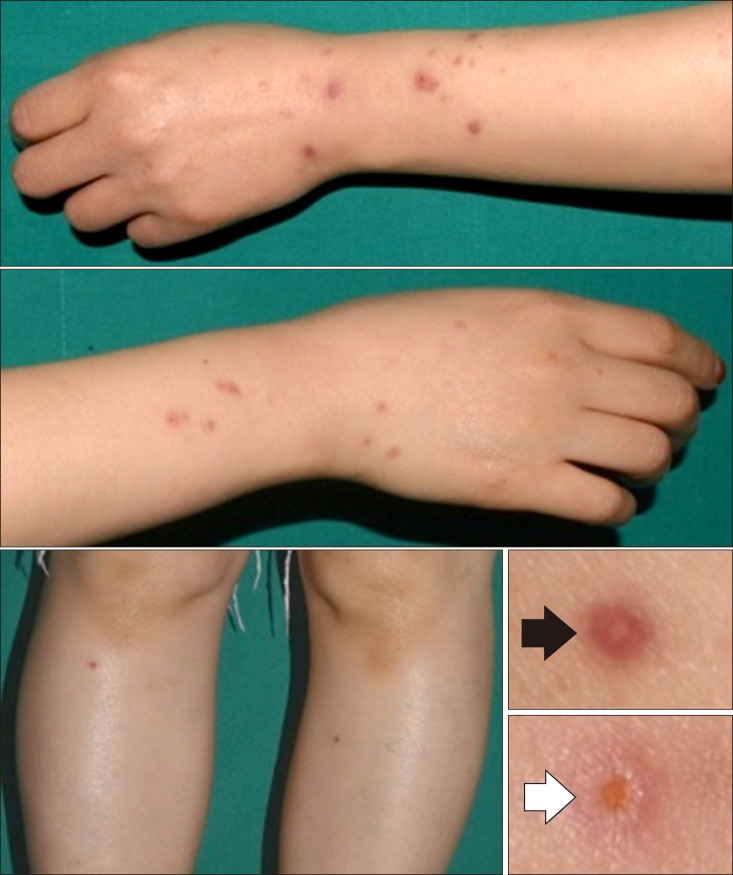
Fig. 1 About 3~5 mm sized, numerous erythematous to brownish colored papules on the extensor surface of limbs. Closer view of the papule shows an umbilication (arrow) and a central crust (empty arrow). We received the patient's consent form about publishing all photographic materials.
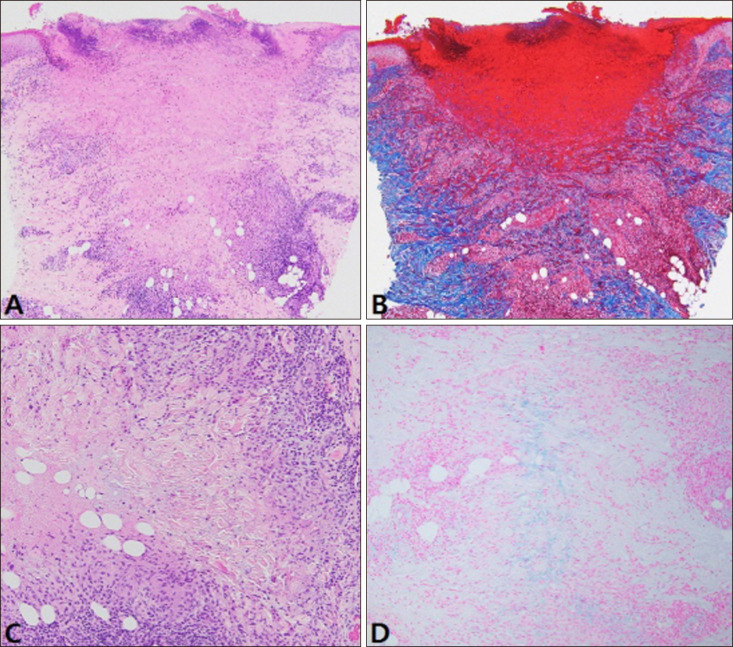
Fig. 2 (A) Wedge-shaped area of necrosis in the upper dermis surrounded by perivascular lymphohistiocytic infiltration (H&E, ×40). (B) The necrotic area is consisted of altered collagen fibers (Masson's trichrome staining, ×40). (C, D) Focal mucin deposition surrounded by palisades of histiocytes near the wedge-shaped dermal necrosis (C: H&E, ×100, D: Alcian blue at pH 2.5, ×100).
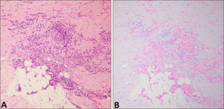
Fig. 3 (A, B) Increased mucin coupled with collection of histiocytes intercalated between collagen bundles in the lower dermis (A: H&E, ×100, B: Alcian blue at pH 2.5, ×100).
Reference
-
1. Piette EW, Rosenbach M. Granuloma annulare: clinical and histologic variants, epidemiology, and genetics. J Am Acad Dermatol. 2016; 75:457–465. PMID: 27543209.2. Tirumalae R, Yeliur IK, Antony M, George G, Kenneth J. Papulonecrotic tuberculid-clinicopathologic and molecular features of 12 Indian patients. Dermatol Pract Concept. 2014; 4:17–22. PMID: 24855568.
Article3. Samlaska CP, Sandberg GD, Maggio KL, Sakas EL. Generalized perforating granuloma annulare. J Am Acad Dermatol. 1992; 27:319–322. PMID: 1517496.
Article4. Penas PF, Jones-Caballero M, Fraga J, Sánchez-Pérez J, García-Díez A. Perforating granuloma annulare. Int J Dermatol. 1997; 36:340–348. PMID: 9199980.
Article5. Seong YK, Cho KH, Kim SH, Lee YS. A case of perforating granuloma annulare. Korean J Dermatol. 1986; 24:678–681.6. Hong KT, Lee KH, Park CI. A case of generalized perforating granuloma annulare. Korean J Dermatol. 1988; 26:560–564.7. Lee JD, Yi JY, Cho BK, Houh W. Anetoderma due to generalized perforating granuloma annulare. Ann Dermatol. 1990; 2:96–99.
Article8. Yoon SY, Kim KH, Suhr KB, Park JK. A case of generalized granuloma annulare with perforating and subcutaneous granuloma annulare. Korean J Dermatol. 1995; 33:1119–1123.9. Lee SY, Han KP, Choi KC, Kim YK. A case of disseminated perforating granuloma annulare in a child. Ann Dermatol. 1996; 8:223–226.
Article10. Lee SW, Lee IW, Lee YS, Lee SC. Generalized perforating granuloma annulare. Korean J Dermatol. 2000; 38:1248–1249.11. Koh BK, Park CJ, Yi JY, Cho BK. A case of generalized perforating granuloma annulare with diabetes mellitus. Korean J Dermatol. 2001; 39:491–493.12. Owens DW, Freeman RG. Perforating granuloma annulare. Arch Dermatol. 1971; 103:64–67. PMID: 5539506.
Article13. Wilson-Jones E, Winkelmann RK. Papulonecrotic tuberculid: a neglected disease in Western countries. J Am Acad Dermatol. 1986; 14:815–826. PMID: 3711385.
Article14. Pereira AR, Vieira MB, Monteiro MP, Enokihara MM, Michalany NS, Bagatin E, et al. Perforating granuloma annulare mimicking papulonecrotic tuberculid. An Bras Dermatol. 2013; 88(6 Suppl 1):101–104. PMID: 24346892.
Article15. Yu JC, Lee KC. Generalised perforating granuloma annulare mimicking papulonecrotic tuberculid. Hong Kong J Dermatol Venereol. 2006; 14:139–142.16. Woo TY, Rasmussen JE. Disorders of transepidermal elimination. Part 2. Int J Dermatol. 1985; 24:337–348. PMID: 3899955.
