Evaluation of Tear Film Lipid Layer Thickness Measurements Obtained Using an Ocular Surface Interferometer in Nasolacrimal Duct Obstruction Patients
- Affiliations
-
- 1Department of Ophthalmology, CHA Bundang Medical Center, CHA University, Seongnam, Korea. eye@cha.ac.kr
- KMID: 2427958
- DOI: http://doi.org/10.3341/kjo.2018.0012
Abstract
- PURPOSE
To compare the tear film lipid layer thickness (LLT) between patients with incomplete nasolacrimal duct obstruction (NLDO) and normal controls and to analyze the changes in tear film LLT and blinking pattern after silicone tube intubation in NLDO patients.
METHODS
We reviewed the medical records of 68 eyes in 52 incomplete NLDO patients who underwent silicone tube intubation from January 2017 to July 2017. The LLT, blinking pattern, and Meibomian gland image were measured with the LipiView II ocular surface interferometer. The Meibomian gland drop-out ratio was measured using the polygon selection tool in the Image J program. Tear meniscus height, which is the other lacrimal indicator, was assessed with spectral-domain optical coherence tomography.
RESULTS
Tear meniscus height was significantly decreased after silicone tube intubation (p < 0.01). Preoperative minimum, maximum, and average LLT values were 62.4 ± 24.0, 86.7 ± 17.9, and 71.7 ± 23.3 nm, respectively. Significant changes in the minimum, maximum, and average LLT (74.8 ± 23.6, 98.8 ± 11.0, and 91.6 ± 16.1 nm, respectively) were observed after silicone tube intubation (p < 0.001, p = 0.001, and p < 0.001). The partial blinking/total blinking ratio in 20 seconds and the Meibomian gland drop-out ratio showed no significant change after silicone tube intubation.
CONCLUSIONS
Overall, the LLT was increased after silicone tube intubation. Silicone tube intubation may be helpful in maintaining LLT with a normalized of amount of tears.
Keyword
MeSH Terms
Figure
-
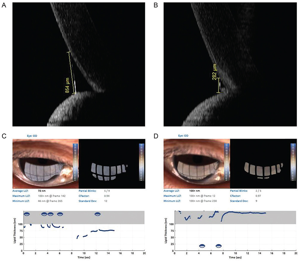
Fig. 1 Tear meniscus height, lipid layer thickness and blinking patterns before and after silicone tube intubation. (A,B) Preoperative and postoperative tear meniscus height obtained using spectral-domain optical coherence tomography (Spectralis, Heidelberg Engineering, Heidelberg, Germany). (C,D) Preoperative and postoperative lipid layer thickness and blinking patterns as measured with LipiView II (TearScience, Morrisville, NC, USA)

Fig. 2 Measurement of Meibomian gland drop-out ratio using the Image J program (National Institutes of Health, Bethesda, MD, USA). Meibomian gland drop-out ratio, ratio of Meibomian gland drop-out area to total area, was measured using a polygonal selection tool of the Image J program.
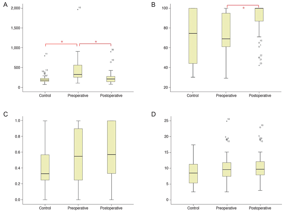
Fig. 3 Box plot of the measured values between control and epiphora patients (before and after silicone tube intubation). (A) Tear meniscus height, (B) lipid layer thickness, (C) partial blinking/total blinking ratio, and (D) Meibomian gland drop-out ratio (*p < 0.05).
Reference
-
1. Mishima S, Maurice DM. The oily layer of the tear film and evaporation from the corneal surface. Exp Eye Res. 1961; 1:39–45.
Article2. Rosenfeld L, Fuller GG. Consequences of interfacial viscoelasticity on thin film stability. Langmuir. 2012; 28:14238–14244.
Article3. Goto E, Tseng SC. Differentiation of lipid tear deficiency dry eye by kinetic analysis of tear interference images. Arch Ophthalmol. 2003; 121:173–180.
Article4. Yokoi N, Yamada H, Mizukusa Y, et al. Rheology of tear film lipid layer spread in normal and aqueous tear-deficient dry eyes. Invest Ophthalmol Vis Sci. 2008; 49:5319–5324.
Article5. Tomlinson A, Doane MG, McFadyen A. Inputs and outputs of the lacrimal system: review of production and evaporative loss. Ocul Surf. 2009; 7:186–198.
Article6. Bron AJ, Tiffany JM, Gouveia SM, et al. Functional aspects of the tear film lipid layer. Exp Eye Res. 2004; 78:347–360.
Article7. Lee JK, Kim TH. Changes in cytokines in tears after endoscopic endonasal dacryocystorhinostomy for primary acquired nasolacrimal duct obstruction. Eye (Lond). 2014; 28:600–607.
Article8. Lew H, Yun YS, Lee SY. Electrolytes and electrophoretic studies of tear proteins in tears of patients with nasolacrimal duct obstruction. Ophthalmologica. 2005; 219:142–146.
Article9. Kubo M, Sakuraba T, Arai Y, Nakazawa M. Tear lipid layer interference changes after dacryocystorhinostomy. Jpn J Ophthalmol. 2001; 45:653–656.
Article10. Munk PL, Lin DT, Morris DC. Epiphora: treatment by means of dacryocystoplasty with balloon dilation of the nasolacrimal drainage apparatus. Radiology. 1990; 177:687–690.
Article11. Sung Y, Park JS, Lew H. Measurement of lacrimal punctum using spectralis domain anterior optical coherence tomography. Acta Ophthalmol. 2017; 95:e619–e624.
Article12. Eom Y, Lee JS, Kang SY, et al. Correlation between quantitative measurements of tear film lipid layer thickness and meibomian gland loss in patients with obstructive meibomian gland dysfunction and normal controls. Am J Ophthalmol. 2013; 155:1104–1110.
Article13. Finis D, Pischel N, Schrader S, Geerling G. Evaluation of lipid layer thickness measurement of the tear film as a diagnostic tool for Meibomian gland dysfunction. Cornea. 2013; 32:1549–1553.
Article14. Arita R, Fukuoka S, Morishige N. new insights into the lipid layer of the tear film and meibomian glands. Eye Contact Lens. 2017; 43:335–339.
Article15. Yokoi N, Takehisa Y, Kinoshita S. Correlation of tear lipid layer interference patterns with the diagnosis and severity of dry eye. Am J Ophthalmol. 1996; 122:818–824.
Article
- Full Text Links
-
- Actions
-
Cited
- CITED
-
- Close
- Share
-
- Similar articles
-
- Evaluation of Changes in Tear Film Lipid Layer Thickness Using Ocular Surface Interferometer after Artificial Tear Application
- Lipid Layer Thickness in Precorneal Tear Film
- Correlation Analysis of Tear Film Lipid Layer Thickness and Ocular Surface Disease Index
- Characteristics of Tear Lipid Layer Patterns on Tearscopy and Lipid Layer Thickness
- Clinical Aspects of Phlyctenular Keratoconjunctivitis Using a Tear Film Interferometer
