Staphylococcal Scalded Skin Syndrome in a Healthy Adult: Easy to Misdiagnose
- Affiliations
-
- 1Department of Plastic and Reconstructive Surgery, Kosin University Gospel Hospital, Kosin University College of Medicine, Busan, Korea. pskwakcy@naver.com
- KMID: 2427394
- DOI: http://doi.org/10.12790/ahm.2018.23.4.271
Abstract
- A 60-year-old male presented with a three-month history of redness and swelling on his left little finger. His medical history was not informative. Wound culture revealed methicillin-resistant Staphylococcus aureus. After vancomycin administration, the skin lesions became worse and whole body bullae and desquamation occurred. This was initially suspected to be a drug eruption; thus, we switched antibiotics from vancomycin to teicoplanin. However, biopsy revealed Staphylococcal scalded skin syndrome (SSSS). After several days, generalized skin symptoms improved. The patient recovered and is in good physical health without recurrence six months later. We describe a localized form of SSSS, which is very rare in healthy adults. Consequently, there is a high risk of misdiagnosis. Thus, we report a rare case of SSSS in a healthy adult and the importance of early histological examination for accurate diagnosis.
MeSH Terms
Figure
-

Fig. 1 Redness and mild swelling on the left little finger.

Fig. 2 There were no radiologic abnormalities.

Fig. 3 Hospital day 4. Despite of 4 days antibiotics treatment, skin symptoms did not improve.

Fig. 4 Hospital day 7. After 3 days of vancomycin use, multiple bullae on the whole body was appeared. (A) Left lateral chest. (B) Back and sacral area.

Fig. 5 Hospital day 12. Skin desquamation and discharge on left hand were even worse. (A) Dorsal side. (B) Palmar aspect.

Fig. 6 Skin biopsy result. Subcorneal splitting of the epidermis with superficial epidermal necrosis is seen. There are few inflammatory cells within the bulla cavity and there is only slight damage to the underlying epidermal cells. It suggests staphylococcal scalded skin syndrome (H&E, ×100).
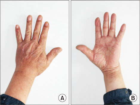
Fig. 7 After discharge from the hospital with a 3-month follow-up clinical photo in the well-healed state. (A) Dorsal side. (B) Palmar aspect.
Reference
-
1. Fitzpatrick TB, Goldsmith LA, Katz SI, Gilchrest BA, Paller AS. Fitzpatrick's dermatology in general medicine. 8th ed. New York: McGraw Hill Medical;2012. p. 2148–2160.2. Miyashita K, Ogawa K, Iioka H, et al. Adult case of Saphylococcal scalded skin syndrome differentiated from toxic epidermal necrolysis with the aid of dermoscopy. J Dermatol. 2016; 43:842–843.3. Aydin D, Alsbjørn B. Severe case of Saphylococcal scalded skin syndrome in a 5-year-old child - case report. Clin Case Rep. 2016; 4:416–419.4. Mishra AK, Yadav P, Mishra A. A systemic review on Staphylococcal scalded skin syndrome (SSSS): a rare and critical disease of neonates. Open Microbiol J. 2016; 10:150–159.
Article5. Cribier B, Piemont Y, Grosshans E. Staphylococcal scalded skin syndrome in adults. A clinical review illustrated with a new case. J Am Acad Dermatol. 1994; 30:319–324.6. Badon HR, King J, Brodell RT, Byrd A. Distinguishing features: Staphylococcal scalded skin syndrome vs toxic epidermal necrolysis. SKIN J Cutan Med. 2018; 2:135–139.
Article7. Patel GK, Finlay AY. Staphylococcal scalded skin syndrome: diagnosis and management. Am J Clin Dermatol. 2003; 4:165–175.8. Ito Y, Funabashi Yoh M, Toda K, Shimazaki M, Nakamura T, Morita E. Staphylococcal scalded-skin syndrome in an adult due to methicillin-resistant Staphylococcal aureus. J Infect Chemother. 2002; 8:256–261.9. Yamaguchi T, Yokota Y, Terajima J, et al. Clonal association of Staphylococcus aureus causing bullous impetigo and the emergence of new methicillin-resistant clonal groups in Kansai district in Japan. J Infect Dis. 2002; 185:1511–1516.
