Left Ventricular Functional Parameters and Geometric Patterns in Korean Adults on Coronary CT Angiography with a 320-Detector-Row CT Scanner
- Affiliations
-
- 1Department of Radiology, College of Medicine, Dong-A University, Busan 49201, Korea.
- 2Department of Cardiology, College of Medicine, Dong-A University, Busan 49201, Korea.
- 3Department of Radiology, Kyungpook National University, Daegu 41944, Korea. jonglee@knu.ac.kr
- KMID: 2427235
- DOI: http://doi.org/10.3348/kjr.2017.18.4.664
Abstract
OBJECTIVE
To assess the normal reference values of left ventricle (LV) functional parameters in Korean adults on coronary CT angiography (CCTA) with a 320-detector-row CT scanner, and to analyze sex-related differences and correlations with various clinical characteristics.
MATERIALS AND METHODS
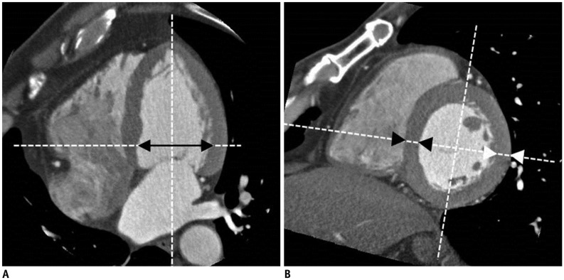
This study retrospectively enrolled 172 subjects (107 men and 65 women; age, 58 ± 10.9 years; body surface area [BSA], 1.75 ± 0.2 m²) who underwent CCTA without any prior history of cardiac disease. The following parameters were measured by post-processing the CT data: LV volume, LV functional parameters (ejection fraction, stroke volume, cardiac output, etc.), LV myocardial mass, LV inner diameter, and LV myocardial thickness (including septal wall thickness [SWT], posterior wall thickness [PWT], and relative wall thickness [RWT = 2 × PWT / LV inner diameter]). All of the functional or volumetric parameters were normalized using the BSA. The general characteristics and co-morbidities for the enrolled subjects were recorded, and the correlations between these factors and the LV parameters were then evaluated.
RESULTS
The LV myocardial thickness (SWT, 1.08 ± 0.18 cm vs. 0.90 ± 0.17 cm, p < 0.001; PWT, 0.91 ± 0.15 cm vs. 0.78 ± 0.10 cm, p < 0.001; RWT, 0.38 ± 0.08 cm vs. 0.33 ± 0.05 cm, p < 0.001), LV volume (LV end-diastolic volume, 112.9 ± 26.1 mL vs. 98.2 ± 21.0 mL, p < 0.001; LV end-systolic volume, 41.7 ± 14.7 mL vs. 33.7 ± 12.2 mL, p = 0.001) and mass (145.0 ± 29.1 g vs. 107.9 ± 20.0 g, p < 0.001) were significantly greater in men than in women. However, these differences were not significant after normalization using BSA, except for the LV mass (LV mass index, 79.6 ± 14.0 g/m² vs. 66.2 ± 11.0 g/m², p < 0.001). The cardiac output and ejection fraction were not significantly different between the men and women (cardiac output, 4.3 ± 1.0 L/min vs. 4.2 ± 0.9 L/min, p = 0.452; ejection fraction, 63.4 ± 7.7% vs. 66.4 ± 7.6%, p = 0.079). Most of the LV parameters were positively correlated with BSA, body weight, and total Agatston score.
CONCLUSION
This study provides sex-related reference values and percentiles for LV on cardiac CT and should assist in interpreting results.
Keyword
MeSH Terms
Figure
Cited by 4 articles
-
Role of Cardiac Computed Tomography in the Diagnosis of Left Ventricular Myocardial Diseases
Sung Min Ko, Sung Ho Hwang, Hye-Jeong Lee
J Cardiovasc Imaging. 2019;27(2):73-92. doi: 10.4250/jcvi.2019.27.e17.Assessment of Left Ventricular Myocardial Diseases with Cardiac Computed Tomography
Sung Min Ko, Tae Hoon Kim, Eun Ju Chun, Jin Young Kim, Sung Ho Hwang
Korean J Radiol. 2019;20(3):333-351. doi: 10.3348/kjr.2018.0280.Age of Data in Contemporary Research Articles Published in Representative General Radiology Journals
Ji Hun Kang, Dong Hwan Kim, Seong Ho Park, Jung Hwan Baek
Korean J Radiol. 2018;19(6):1172-1178. doi: 10.3348/kjr.2018.19.6.1172.Clinical Applications of Wide-Detector CT Scanners for Cardiothoracic Imaging: An Update
Eun-Ju Kang
Korean J Radiol. 2019;20(12):1583-1596. doi: 10.3348/kjr.2019.0327.
Reference
-
1. Raff GL, Gallagher MJ, O'Neill WW, Goldstein JA. Diagnostic accuracy of noninvasive coronary angiography using 64-slice spiral computed tomography. J Am Coll Cardiol. 2005; 46:552–557. PMID: 16053973.
Article2. Kim YJ, Yong HS, Kim SM, Kim JA, Yang DH, Hong YJ, et al. Korean guidelines for the appropriate use of cardiac CT. Korean J Radiol. 2015; 16:251–285. PMID: 25741189.
Article3. Lin FY, Min JK. Assessment of cardiac volumes by multidetector computed tomography. J Cardiovasc Comput Tomogr. 2008; 2:256–262. PMID: 19083959.
Article4. Mahnken AH, Bruners P, Schmidt B, Bornikoel C, Flohr T, Günther RW. Left ventricular function can reliably be assessed from dual-source CT using ECG-gated tube current modulation. Invest Radiol. 2009; 44:384–389. PMID: 19448556.
Article5. Juergens KU, Grude M, Maintz D, Fallenberg EM, Wichter T, Heindel W, et al. Multi-detector row CT of left ventricular function with dedicated analysis software versus MR imaging: initial experience. Radiology. 2004; 230:403–410. PMID: 14668428.
Article6. Raman SV, Shah M, McCarthy B, Garcia A, Ferketich AK. Multi-detector row cardiac computed tomography accurately quantifies right and left ventricular size and function compared with cardiac magnetic resonance. Am Heart J. 2006; 151:736–744. PMID: 16504643.
Article7. Asferg C, Usinger L, Kristensen TS, Abdulla J. Accuracy of multi-slice computed tomography for measurement of left ventricular ejection fraction compared with cardiac magnetic resonance imaging and two-dimensional transthoracic echocardiography: a systematic review and meta-analysis. Eur J Radiol. 2012; 81:e757–e762. PMID: 22381439.8. Stolzmann P, Scheffel H, Leschka S, Schertler T, Frauenfelder T, Kaufmann PA, et al. Reference values for quantitative left ventricular and left atrial measurements in cardiac computed tomography. Eur Radiol. 2008; 18:1625–1634. PMID: 18446346.
Article9. Lin FY, Devereux RB, Roman MJ, Meng J, Jow VM, Jacobs A, et al. Cardiac chamber volumes, function, and mass as determined by 64-multidetector row computed tomography: mean values among healthy adults free of hypertension and obesity. JACC Cardiovasc Imaging. 2008; 1:782–786. PMID: 19356515.10. Gardin JM, Wagenknecht LE, Anton-Culver H, Flack J, Gidding S, Kurosaki T, et al. Relationship of cardiovascular risk factors to echocardiographic left ventricular mass in healthy young black and white adult men and women. The CARDIA study. Coronary Artery Risk Development in Young Adults. Circulation. 1995; 92:380–387. PMID: 7634452.11. Natori S, Lai S, Finn JP, Gomes AS, Hundley WG, Jerosch-Herold M, et al. Cardiovascular function in multi-ethnic study of atherosclerosis: normal values by age, sex, and ethnicity. AJR Am J Roentgenol. 2006; 186(6 Suppl 2):S357–S365. PMID: 16714609.
Article12. Chen JJ, Manning MA, Frazier AA, Jeudy J, White CS. CT angiography of the cardiac valves: normal, diseased, and postoperative appearances. Radiographics. 2009; 29:1393–1412. PMID: 19755602.
Article13. Lang RM, Bierig M, Devereux RB, Flachskampf FA, Foster E, Pellikka PA, et al. Recommendations for chamber quantification. Eur J Echocardiogr. 2006; 7:79–108. PMID: 16458610.
Article14. Lang RM, Badano LP, Mor-Avi V, Afilalo J, Armstrong A, Ernande L, et al. Recommendations for cardiac chamber quantification by echocardiography in adults: an update from the American Society of Echocardiography and the European Association of Cardiovascular Imaging. Eur Heart J Cardiovasc Imaging. 2015; 16:233–270. PMID: 25712077.
Article15. Christner JA, Kofler JM, McCollough CH. Estimating effective dose for CT using dose-length product compared with using organ doses: consequences of adopting International Commission on Radiological Protection publication 103 or dual-energy scanning. AJR Am J Roentgenol. 2010; 194:881–889. PMID: 20308486.
Article16. Evans JD. Straightforward statistics for the behavioral sciences. Pacific Grove: Brooks/Cole Publishing;1996.17. Nham E, Kim SM, Lee SC, Chang SA, Sung J, Cho SJ, et al. Association of cardiovascular disease risk factors with left ventricular mass, biventricular function, and the presence of silent myocardial infarction on cardiac MRI in an asymptomatic population. Int J Cardiovasc Imaging. 2016; 32(Suppl 1):173–181.
Article18. Bernard Y, Meneveau N, Boucher S, Magnin D, Anguenot T, Schiele F, et al. Lack of agreement between left ventricular volumes and ejection fraction determined by two-dimensional echocardiography and contrast cineangiography in postinfarction patients. Echocardiography. 2001; 18:113–122. PMID: 11262534.
Article19. Picard MH, Popp RL, Weyman AE. Assessment of left ventricular function by echocardiography: a technique in evolution. J Am Soc Echocardiogr. 2008; 21:14–21. PMID: 18165124.
Article20. Chang SA, Choe YH, Jang SY, Kim SM, Lee SC, Oh JK. Assessment of left and right ventricular parameters in healthy Korean volunteers using cardiac magnetic resonance imaging: change in ventricular volume and function based on age, gender and body surface area. Int J Cardiovasc Imaging. 2012; 28(Suppl 2):141–147. PMID: 23139150.
Article21. Sandstede J, Lipke C, Beer M, Hofmann S, Pabst T, Kenn W, et al. Age- and gender-specific differences in left and right ventricular cardiac function and mass determined by cine magnetic resonance imaging. Eur Radiol. 2000; 10:438–442.
Article
- Full Text Links
- Actions
-
Cited
- CITED
-
- Close
- Share
- Similar articles
-
- Unusual Coronary Artery Fistula: Left Anterior Descending Coronary Artery - Left Ventricular Fistula Diagnosed by ECG-Gated Multi-Detector Row Coronary CT Angiography
- Clinical Applications of Wide-Detector CT Scanners for Cardiothoracic Imaging: An Update
- 16-Slice Multi-Detector Row CT Coronary Angiography: Image Quality and Optimization of the Image Reconstruction Window
- Normal coronary CT angiography with subsequent adverse cardiac events
- MDCT Application in the Vascular System


