Diagnostic Clue of Meningeal Melanocytoma: Case Report and Review of Literature
- Affiliations
-
- 1Department of Neurosurgery, Asan Medical Center, University of Ulsan College of Medicine, Seoul, Korea. scrhim@amc.seoul.kr
- 2Department of Radiology, Asan Medical Center, University of Ulsan College of Medicine, Seoul, Korea.
- KMID: 2427140
- DOI: http://doi.org/10.3349/ymj.2017.58.2.467
Abstract
- In this report, the patient was pre-diagnosed as meningioma before surgery, which turned out to be meningeal melanocytoma. Hence, we will discuss the interpretation of imaging and neurological statuses that may help avoid this problem. A 45-year-old man had increasing pain around the neck 14 months prior to admission. His cervical spine MR imaging revealed a space-occupying, contrast-enhancing mass within the dura at the level of C1. The neurologic examination revealed that the patient had left-sided lower extremity weakness of 4+, decreased sensation on the right side, and hyperreflexia in both legs. Department of Neuroradiology interpreted CT and MR imaging as meningiom. The patient underwent decompression and removal of the mass. We confirmed diagnosis as meningeal melanocytoma through pathologic findings. Afterwards, we reviewed the patient's imaging work-up, which showed typical findings of meningeal melanocytoma. However, it was mistaken as meningioma, since the disease is rare.
Keyword
MeSH Terms
Figure
-
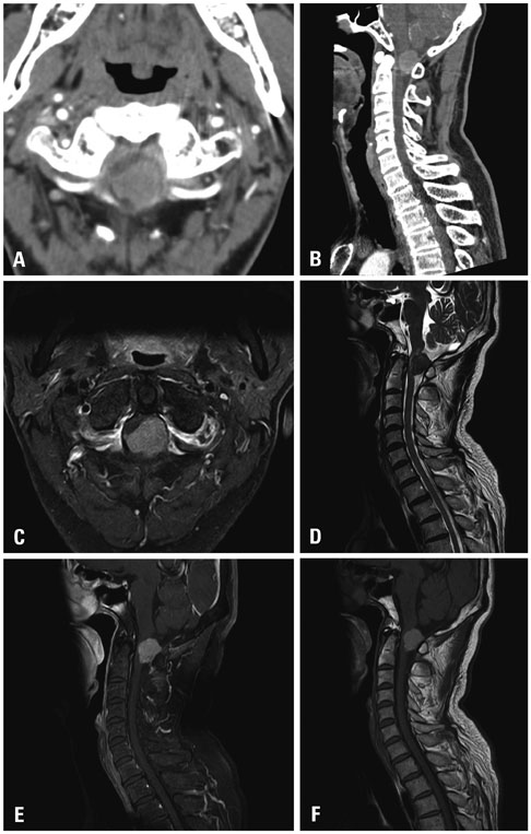
Fig. 1 An approximately 3-cm T1 high signal intensity and T2 low signal intensity homogenous enhancing intradural extramedullary mass abutting the left posterolateral aspect of the dura at C1 level, without adjacent dural thickening or dural tail sign, no evidence of abnormal bone change. (A) Contrast CT axial image. (B) Contrast CT saggital image. (C) T1-enhanced image. (D) T2 sagittal image. (E) T1 enhanced sagittal image. (F) T1 sagittal image.
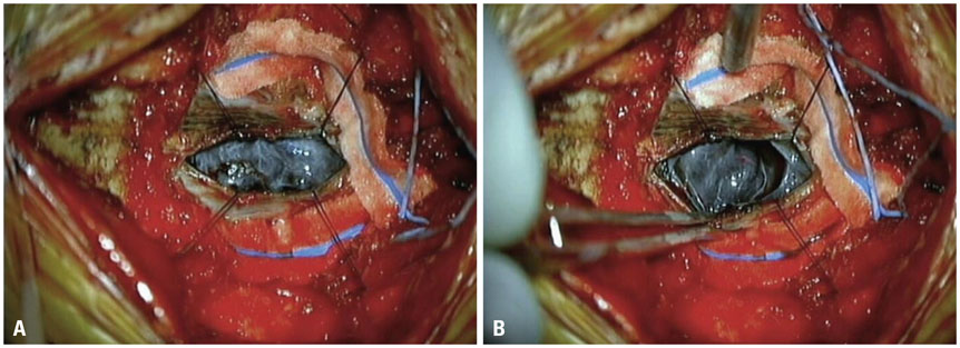
Fig. 2 (A and B) Intraoperative microscopic findings. A black colored, well-circumscribed, nodular tumor was seen. The tumor was a soft mass and well capsulated.

Fig. 3 The results of immunohistochemical staining. (A) Hematoxylin and eosin staining (×100). (B) HMB45 staining (×20). (C) S-100 protein staining (×20). (D) Ki-67 labeling index (<5%) (×100). Microscopic features supporting malignant melanoma, such as mitoses, necrosis, or nuclear pleomorphism were not observed, and the Ki-67 labeling index was low.
Reference
-
1. Ahluwalia S, Ashkan K, Casey AT. Meningeal melanocytoma: clinical features and review of the literature. Br J Neurosurg. 2003; 17:347–351.
Article2. Limas C, Tio FO. Meningeal melanocytoma (“melanotic meningioma”). Its melanocytic origin as revealed by electron microscopy. Cancer. 1972; 30:1286–1294.
Article3. Bhargava P, Grewal SS, Dewan Y, Jhawar SS, Jain V, Gupta B. et al. Craniovertebral junction melanocytoma: a case report. Turk Neurosurg. 2013; 23:539–542.4. Clarke DB, Leblanc R, Bertrand G, Quartey GR, Snipes GJ. Meningeal melanocytoma. Report of a case and a historical comparison. J Neurosurg. 1998; 88:116–121.5. Hou GQ, Sun JC, Zhang XJ, Shen BX, Zhu XJ, Liang L, et al. MR imaging findings of the intraspinal meningeal melanocytoma: correlation with histopathologic findings. AJNR Am J Neuroradiol. 2012; 33:1525–1529.
Article6. Brat DJ, Giannini C, Scheithauer BW, Burger PC. Primary melanocytic neoplasms of the central nervous systems. Am J Surg Pathol. 1999; 23:745–754.7. Liubinas SV, Maartens N, Drummond KJ. Primary melanocytic neoplasms of the central nervous system. J Clin Neurosci. 2010; 17:1227–1232.
Article8. Litofsky NS, Zee CS, Breeze RE, Chandrasoma PT. Meningeal melanocytoma: diagnostic criteria for a rare lesion. Neurosurgery. 1992; 31:945–948.9. Uematsu Y, Yukawa S, Yokote H, Itakura T, Hayashi S, Komai N. Meningeal melanocytoma: magnetic resonance imaging characteristics and pathological features. Case report. J Neurosurg. 1992; 76:705–709.10. Sealy RC. Radicals in melanin biochemistry. Methods Enzymol. 1984; 105:479–483.11. Sen R, Sethi D, Goyal V, Duhan A, Modi S. Spinal meningeal melanocytoma. Asian J Neurosurg. 2011; 6:110–112.
Article12. Beseoglu K, Knobbe CB, Reifenberger G, Steiger HJ, Stummer W. Supratentorial meningeal melanocytoma mimicking a convexity meningioma. Acta Neurochir (Wien). 2006; 148:485–490.
Article
- Full Text Links
-
- Actions
-
Cited
- CITED
-
- Close
- Share
-
- Similar articles
-
- Primary Spinal Meningeal Melanocytoma
- Meningeal Melanocytoma Associated with Ota's Nevus: Report of a case
- Primary Meningeal Melanocytoma in the Thoracic Spine: A Case Report
- Primary Intramedullary Meningeal Melanocytoma in Cervical Spine: A Case Report and Literature Review
- Spinal Meningeal Melanocytoma with Benign Histology Showing Leptomeningeal Spread: Case Report
