Korean J Radiol.
2013 Jun;14(3):470-476. 10.3348/kjr.2013.14.3.470.
Spinal Meningeal Melanocytoma with Benign Histology Showing Leptomeningeal Spread: Case Report
- Affiliations
-
- 1Department of Radiology, Inje University Haeundae Paik Hospital, Busan 612-030, Korea. okkimmd@hanafos.com
- 2Department of Radiology, Inje University Busan Paik Hospital, Busan 633-165, Korea.
- 3Department of Radiology, Busan National University Hospital, Busan 602-739, Korea.
- 4Department of Pathology, Inje University Haeundae Paik Hospital, Busan 612-030, Korea.
- 5Department of Neurosurgery, Inje University Haeundae Paik Hospital, Busan 612-030, Korea.
- KMID: 1705462
- DOI: http://doi.org/10.3348/kjr.2013.14.3.470
Abstract
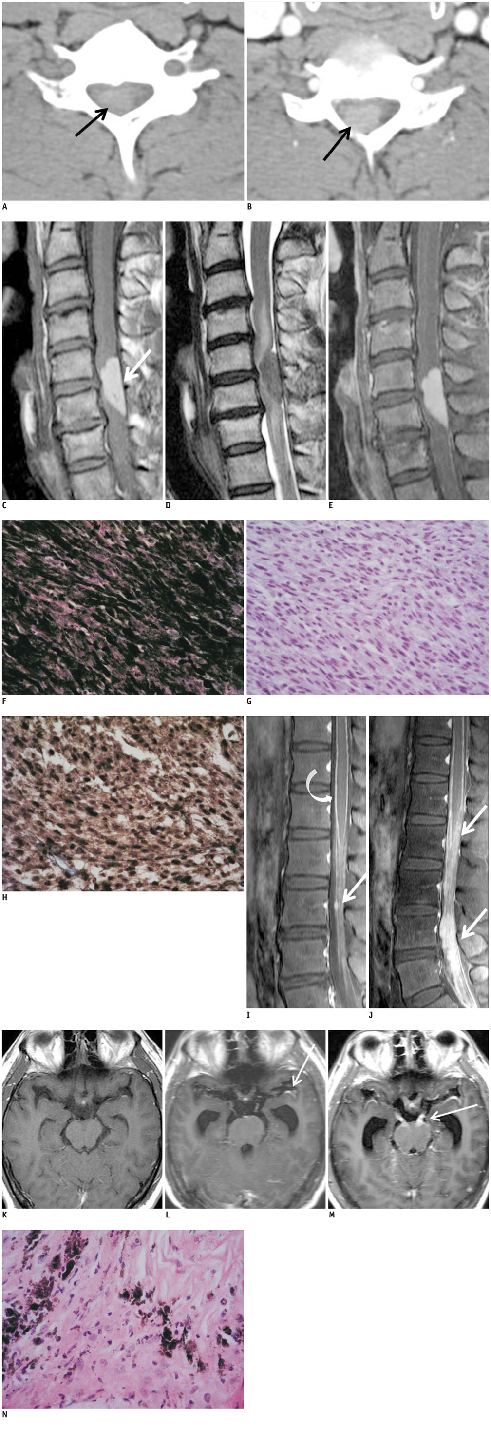
- Meningeal melanocytoma is a rare benign tumor with relatively good prognosis. However, local aggressive behavior of meningeal melanocytoma has been reported, especially in cases of incomplete surgical resection. Malignant transformation was raised as possible cause by prior reports to explain this phenomenon. We present an unusual case of meningeal melanocytoma associated with histologically benign leptomeningeal spread and its subsequent aggressive clinical course, and describe its radiological findings.
Keyword
MeSH Terms
Figure
Reference
-
1. Painter TJ, Chaljub G, Sethi R, Singh H, Gelman B. Intracranial and intraspinal meningeal melanocytosis. AJNR Am J Neuroradiol. 2000. 21:1349–1353.2. Roser F, Nakamura M, Brandis A, Hans V, Vorkapic P, Samii M. Transition from meningeal melanocytoma to primary cerebral melanoma. Case report. J Neurosurg. 2004. 101:528–531.3. Wang F, Li X, Chen L, Pu X. Malignant transformation of spinal meningeal melanocytoma. Case report and review of the literature. J Neurosurg Spine. 2007. 6:451–454.4. Uozumi Y, Kawano T, Kawaguchi T, Kaneko Y, Ooasa T, Ogasawara S, et al. Malignant transformation of meningeal melanocytoma: a case report. Brain Tumor Pathol. 2003. 20:21–25.5. Bydon A, Gutierrez JA, Mahmood A. Meningeal melanocytoma: an aggressive course for a benign tumor. J Neurooncol. 2003. 64:259–263.6. Limas C, Tio FO. Meningeal melanocytoma ("melanotic meningioma"). Its melanocytic origin as revealed by electron microscopy. Cancer. 1972. 30:1286–1294.7. Chacko G, Rajshekhar V. Thoracic intramedullary melanocytoma with long-term follow-up. J Neurosurg Spine. 2008. 9:589–592.8. Chen CJ, Hsu YI, Ho YS, Hsu YH, Wang LJ, Wong YC. Intracranial meningeal melanocytoma: CT and MRI. Neuroradiology. 1997. 39:811–814.9. Brat DJ. Perry A, Brat DJ, editors. Melanocytic Neoplasm of Central Nerveous System. Practical Surgical Neuropathology: A Diagnostic Approach. 2010. Philadelphia: Churchill Livingstone;353–359.10. Clarke DB, Leblanc R, Bertrand G, Quartey GR, Snipes GJ. Meningeal melanocytoma. Report of a case and a historical comparison. J Neurosurg. 1998. 88:116–121.
- Full Text Links
- Actions
-
Cited
- CITED
-
- Close
- Share
- Similar articles
-
- Primary Spinal Meningeal Melanocytoma
- Primary Meningeal Melanocytoma in the Thoracic Spine: A Case Report
- Meningeal Melanocytoma Associated with Ota's Nevus: Report of a case
- Spinal meningeal melanocytoma
- Primary Intramedullary Meningeal Melanocytoma in Cervical Spine: A Case Report and Literature Review


