Yonsei Med J.
2017 Mar;58(2):458-461. 10.3349/ymj.2017.58.2.458.
Coronary Stent Infection Presented as Recurrent Stent Thrombosis
- Affiliations
-
- 1Division of Interventional Cardiology, Cardiovascular Center, Taichung Veterans General Hospital, Taichung, Taiwan, R.O.C. wcchangvghtc@gmail.com
- 2School of Medicine, National Yang-Ming University, Taipei, Taiwan, R.O.C.
- 3Division of Cardiovascular Surgery, Department of Surgery, Feng Yuan Hospital, Taichung, Taiwan, R.O.C.
- KMID: 2427138
- DOI: http://doi.org/10.3349/ymj.2017.58.2.458
Abstract
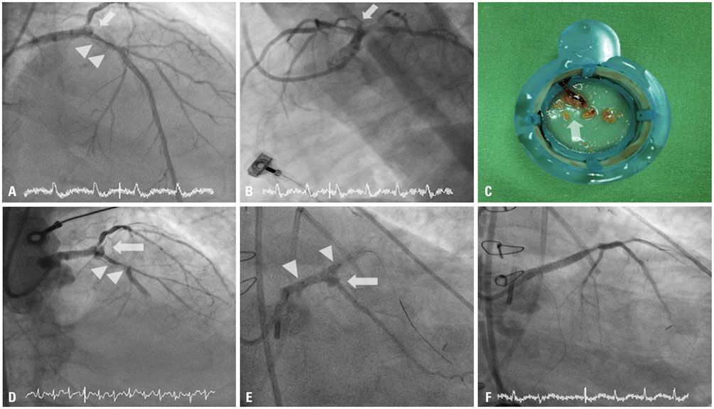
- Percutaneous transluminal coronary angioplasty with metal stent placement has become a well-developed treatment modality for coronary stenotic lesions. Although infection involving implanted stents is rare, it can, however, occur with high morbidity and mortality. We describe herein a case of an inserted coronary stent that was infected and complicated with recurrent stent thrombosis, pseudoaneurysm formation and severe sepsis. Despite repeated intervention and bypass surgery, the patient died from severe sepsis.
MeSH Terms
Figure
Reference
-
1. Bosman WM, Borger van der Burg BL, Schuttevaer HM, Thoma S, Hedeman Joosten PP. Infections of intravascular bare metal stents: a case report and review of literature. Eur J Vasc Endovasc Surg. 2014; 47:87–99.
Article2. Roubelakis A, Rawlins J, Baliulis G, Olsen S, Corbett S, Kaarne M, et al. Coronary artery rupture caused by stent infection: a rare complication. Circulation. 2015; 131:1302–1303.3. Wu EB, Chan WW, Yu CM. Left main stem rupture caused by methicillin resistant Staphylococcus aureus infection of left main stent treated by covered stenting. Int J Cardiol. 2010; 144:e39–e41.
Article4. Garg RK, Sear JE, Hockstad ES. Spontaneous coronary artery perforation secondary to a sirolimus-eluting stent infection. J Invasive Cardiol. 2007; 19:E303–E306.
- Full Text Links
- Actions
-
Cited
- CITED
-
- Close
- Share
- Similar articles
-
- Recurrent Stent Thrombosis in Different Coronary Arteries
- Coincidental Occurrence of Acute In-stent Thrombosis and Iatrogenic Vessel Perforation During a Wingspan Stent Placement: Management with a Stent In-stent Technique
- Recurrent Very Late Stent Thrombosis in a Systemic Lupus Erythematous Patient
- Risk of Stent Stenosis after Implanting a First-Generation Drug-Eluting Stent and Drug Balloon Angioplasty
- Drug-Eluting Stent Used to Treat a Case of Recurrent Right Coronary Artery In-Stent Restenoses often Accompanied by Acute Inferior Wall Myocardial Infarction


