Korean J Orthod.
2016 Nov;46(6):372-378. 10.4041/kjod.2016.46.6.372.
Comparison of postoperative changes in the distal and proximal segments between conventional and sliding mini-plate fixation following mandibular setback
- Affiliations
-
- 1Department of Orthodontics, Dental Research Institute, Pusan National University Dental Hospital, Yangsan, Korea. kimyongil@pusan.ac.kr
- 2Department of Orthodontics, School of Dentistry, University of North Carolina, Chapel Hill, NC, USA.
- 3Department of Orthodontics, Biomedical Research Institute, Pusan National University Hospital, Busan, Korea.
- 4Institute of Translational Dental Sciences, Pusan National University, Busan, Korea.
- KMID: 2426681
- DOI: http://doi.org/10.4041/kjod.2016.46.6.372
Abstract
OBJECTIVE
The purpose of the present study was to evaluate the postoperative three-dimensional (3D) changes in the proximal segments after mandibular setback sagittal split ramus osteotomy and to compare the changes between the conventional mini-plate fixation and semi-rigid sliding plate fixation.
METHODS
Cone-beam computed tomography (CBCT) images were used to evaluate the postoperative 3D changes in the proximal segments during the healing process. CBCT images were superimposed using the symphysis and the lower anterior mandible as references.
RESULTS
There were no statistically significant differences between the conventional mini-plate and semi-rigid sliding plate groups (p > 0.05). With respect to the distribution of changes greater than 2 mm in the landmarks, the right condylion, right coronoid process, and left condylion showed ratios of 55.6%, 50.0%, and 44.4%, respectively, in the semi-rigid sliding plate group; however, none of the landmarks showed ratios greater than 30% in the conventional mini-plate group.
CONCLUSIONS
There were no statistically significant differences in postoperative changes in the segments between the conventional mini-plate and semi-rigid sliding plate groups. Nevertheless, while selecting the type of fixation technique, clinicians should consider that landmarks with greater than 2 mm changes were higher in the semi-rigid sliding plate group than in the conventional mini-plate group.
Figure
-
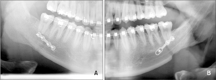
Figure 1 Two different fixation methods. A, Rigid fixation with a mini-plate; B, semi-rigid fixation with a sliding mini-plate.
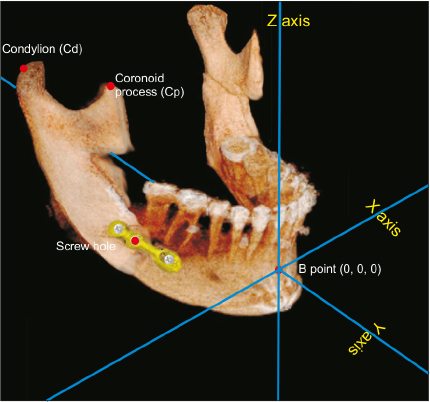
Figure 2 The X, Y, and Z coordinates and landmarks on a cone-beam computed tomography image.
Reference
-
1. Dolce C, Van Sickels JE, Bays RA, Rugh JD. Skeletal stability after mandibular advancement with rigid versus wire fixation. J Oral Maxillofac Surg. 2000; 58:1219–1227.
Article2. Mobarak KA, Krogstad O, Espeland L, Lyberg T. Long-term stability of mandibular setback surgery: a follow-up of 80 bilateral sagittal split osteotomy patients. Int J Adult Orthodon Orthognath Surg. 2000; 15:83–95.3. Costa F, Robiony M, Sembronio S, Polini F, Politi M. Stability of skeletal Class III malocclusion after combined maxillary and mandibular procedures. Int J Adult Orthodon Orthognath Surg. 2001; 16:179–192.4. Kim MJ, Kim SG, Park YW. Positional stability following intentional posterior ostectomy of the distal segment in bilateral sagittal split ramus osteotomy for correction of mandibular prognathism. J Craniomaxillofac Surg. 2002; 30:35–40.
Article5. Borstlap WA, Stoelinga PJ, Hoppenreijs TJ, van't Hof MA. Stabilisation of sagittal split advancement osteotomies with miniplates: a prospective, multicentre study with two-year follow-up. Part III--condylar remodelling and resorption. Int J Oral Maxillofac Surg. 2004; 33:649–655.
Article6. Politi M, Costa F, Cian R, Polini F, Robiony M. Stability of skeletal class III malocclusion after combined maxillary and mandibular procedures: rigid internal fixation versus wire osteosynthesis of the mandible. J Oral Maxillofac Surg. 2004; 62:169–181.
Article7. Rhee CH, Choi YK, Kim YI, Kim SS, Park SB, Son WS. Correlation between skeletal and dental changes after mandibular setback surgery-first orthodontic treatment: Cone-beam computed tomography-generated half-cephalograms. Korean J Orthod. 2015; 45:59–65.
Article8. Proffit WR, Phillips C, Dann C 4th, Turvey TA. Stability after surgical-orthodontic correction of skeletal Class III malocclusion. I. Mandibular setback. Int J Adult Orthodon Orthognath Surg. 1991; 6:7–18.9. Joss CU, Vassalli IM. Stability after bilateral sagittal split osteotomy setback surgery with rigid internal fixation: a systematic review. J Oral Maxillofac Surg. 2008; 66:1634–1643.
Article10. Wolford LM. Concomitant temporomandibular joint and orthognathic surgery. J Oral Maxillofac Surg. 2003; 61:1198–1204.
Article11. Son S, Kim SS, Son WS, Kim YI, Kim YD, Shin SH. Miniscrews versus surgical archwires for intermaxillary fixation in adults after orthognathic surgery. Korean J Orthod. 2015; 45:3–12.
Article12. Law JH, Rotskoff KS, Smith RJ. Stability following combined maxillary and mandibular osteotomies treated with rigid internal fixation. J Oral Maxillofac Surg. 1989; 47:128–136.
Article13. Stroster TG, Pangrazio-Kulbersh V. Assessment of condylar position following bilateral sagittal split ramus osteotomy with wire fixation or rigid fixation. Int J Adult Orthodon Orthognath Surg. 1994; 9:55–63.14. Kim YI, Park SB, Jung YH, Hwang DS, Lee JY. Evaluation of intersegmental displacement according to osteosynthesis method for mandibular setback sagittal split ramus osteotomy using cone-beam computed tomographic superimposition. J Oral Maxillofac Surg. 2012; 70:2893–2898.
Article15. Mavili ME, Canter HI, Saglam-Aydinatay B. Semirigid fixation of mandible and maxilla in orthognathic surgery: stability and advantages. Ann Plast Surg. 2009; 63:396–403.16. Baek RM, Lee SW. A new condyle repositionable plate for sagittal split ramus osteotomy. J Craniofac Surg. 2010; 21:489–490.
Article17. Proffit WR, Phillips C, Turvey TA. Stability after mandibular setback: mandible-only versus 2-jaw surgery. J Oral Maxillofac Surg. 2012; 70:e408–e414.
Article18. Korkmaz HH. Evaluation of different miniplates in fixation of fractured human mandible with the finite element method. Oral Surg Oral Med Oral Pathol Oral Radiol Endod. 2007; 103:e1–e13.
Article19. Tams J, van Loon JP, Otten E, Rozema FR, Bos RR. A three-dimensional study of bending and torsion moments for different fracture sites in the mandible: an in vitro study. Int J Oral Maxillofac Surg. 1997; 26:383–388.
Article
- Full Text Links
-
- Actions
-
Cited
- CITED
-
- Close
- Share
-
- Similar articles
-
- Comparative Study of Skeletal Relapse According to the Fixation Method after BSSRO for Mandibular Setback
- Skeletal relapse and dental change during intermaxillary fixation after mandibular setback
- Cephalometric study on the effect caused by different fixation plate removal time on the early skeletal relapse
- Mini-T Plate Fixation for Neer Type II Distal Clavicle Fracture
- Rupture of the Extensor Pollicis Longus Tendon at the Proximal Screw of Volar Plate Fixation for Distal Radius Fracture: A Case Report
