Clin Orthop Surg.
2018 Dec;10(4):462-467. 10.4055/cios.2018.10.4.462.
Morphometric Variations in the Volar Aspect of the Distal Radius
- Affiliations
-
- 1Department of Orthopedic Surgery, Hallym University Sacred Heart Hospital, Anyang, Korea. cartilage11@hotmail.com
- 2Jeil Medical Corporation, Seoul, Korea.
- KMID: 2426532
- DOI: http://doi.org/10.4055/cios.2018.10.4.462
Abstract
- BACKGROUND
Significant discrepancy exists between anatomical plate designs and the anatomy of the native distal radius, which may be attributable to considerable morphometric variations in the volar aspect of the distal radius. We aimed to evaluate the degree of variability in the morphometry of the distal radius and identify factors associated with this variability.
METHODS
We measured the volar surface angle (VSA) of the intermediate and lateral columns and the volar surface width (VSW) in the distal radius from three-dimensional computed tomography scans acquired from 81 cadaveric forearms. These morphometric parameters were compared between the lateral and intermediate columns, between males and females, and between Koreans and Caucasians. Caucasian morphometric data were obtained and pooled from the previous studies. The coefficient of variation was used to assess the variability of the parameters and Cohen's d to estimate the effect size of the difference between groups.
RESULTS
The average VSA of the lateral column was 22°± 6°, and that of the intermediate column was 29°± 8° in Koreans (p < 0.001). The variability was high for both VSAs. The VSA of the intermediate column was significantly larger in males than in females (p < 0.001) and in Caucasians than in Koreans (p < 0.001). The average VSW of distal radius was 30 ± 3 mm at the watershed line, and it became narrower proximally. The VSW was significantly larger in males than in females (p < 0.001) and in Koreans than in Caucasians (p < 0.001). The effect sizes of the difference for the VSA and VSW between sexes, races and columns were medium to large.
CONCLUSIONS
Considerable variability exists in the morphometry of the volar distal radius, with sex, race, and column as contributing factors. These results suggest that surgeons should carefully choose an anatomical volar locking plate with appropriate angulation characteristics for each patient to achieve patient-specific alignment of the distal radius.
Keyword
Figure
-
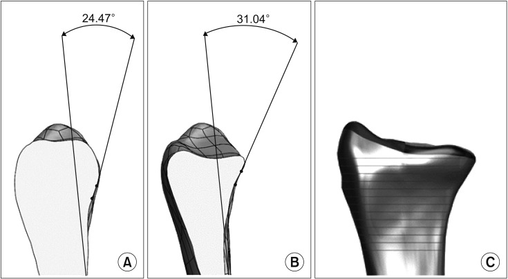
Fig. 1 Measurement methods of the volar surface angle (VSA) at the lateral column and the intermediate column, as well as the volar surface width (VSW) are shown. (A) The VSA at the lateral column is defined as the angle formed by the line along the volar surface of the radial shaft and the line along the volar surface of the radial metaphysis at the center of the scaphoid fossa. (B) The VSA at the intermediate column is defined as the angle formed by the lines drawn in the same fashion at the center of the lunate fossa. (C) The VSW is defined as the length of the line drawn from the lateral to the medial margin of the volar surface of the distal radius, perpendicular to the central axis of the radial shaft.
Reference
-
1. Rikli DA, Regazzoni P. Fractures of the distal end of the radius treated by internal fixation and early function: a preliminary report of 20 cases. J Bone Joint Surg Br. 1996; 78(4):588–592. PMID: 8682826.2. Jakob M, Rikli DA, Regazzoni P. Fractures of the distal radius treated by internal fixation and early function: a prospective study of 73 consecutive patients. J Bone Joint Surg Br. 2000; 82(3):340–344. PMID: 10813166.3. Orbay JL, Fernandez DL. Volar fixation for dorsally displaced fractures of the distal radius: a preliminary report. J Hand Surg Am. 2002; 27(2):205–215. PMID: 11901379.
Article4. Chung KC, Shauver MJ, Birkmeyer JD. Trends in the United States in the treatment of distal radial fractures in the elderly. J Bone Joint Surg Am. 2009; 91(8):1868–1873. PMID: 19651943.
Article5. Richard MJ, Wartinbee DA, Riboh J, Miller M, Leversedge FJ, Ruch DS. Analysis of the complications of palmar plating versus external fixation for fractures of the distal radius. J Hand Surg Am. 2011; 36(10):1614–1620. PMID: 21849236.
Article6. Sammer DM, Fuller DS, Kim HM, Chung KC. A comparative study of fragment-specific versus volar plate fixation of distal radius fractures. Plast Reconstr Surg. 2008; 122(5):1441–1450. PMID: 18971728.
Article7. Osada D, Kamei S, Masuzaki K, Takai M, Kameda M, Tamai K. Prospective study of distal radius fractures treated with a volar locking plate system. J Hand Surg Am. 2008; 33(5):691–700. PMID: 18590852.
Article8. Wei DH, Raizman NM, Bottino CJ, Jobin CM, Strauch RJ, Rosenwasser MP. Unstable distal radial fractures treated with external fixation, a radial column plate, or a volar plate: a prospective randomized trial. J Bone Joint Surg Am. 2009; 91(7):1568–1577. PMID: 19571078.9. Mignemi ME, Byram IR, Wolfe CC, et al. Radiographic outcomes of volar locked plating for distal radius fractures. J Hand Surg Am. 2013; 38(1):40–48. PMID: 23218558.
Article10. Knudsen R, Bahadirov Z, Damborg F. High rate of complications following volar plating of distal radius fractures. Dan Med J. 2014; 61(10):A4906. PMID: 25283615.11. Jones CW, Lawson RD. One size does not fit all: distal radioulnar joint dysfunction after volar locking plate fixation. J Wrist Surg. 2014; 3(1):42–45. PMID: 24533245.12. Casaletto JA, Machin D, Leung R, Brown DJ. Flexor pollicis longus tendon ruptures after palmar plate fixation of fractures of the distal radius. J Hand Surg Eur Vol. 2009; 34(4):471–474. PMID: 19395539.
Article13. Soong M, Earp BE, Bishop G, Leung A, Blazar P. Volar locking plate implant prominence and flexor tendon rupture. J Bone Joint Surg Am. 2011; 93(4):328–335. PMID: 21239658.
Article14. Buzzell JE, Weikert DR, Watson JT, Lee DH. Precontoured fixed-angle volar distal radius plates: a comparison of anatomic fit. J Hand Surg Am. 2008; 33(7):1144–1152. PMID: 18762111.
Article15. Oppermann J, Wacker M, Stein G, et al. Anatomical fit of seven different palmar distal radius plates. Arch Orthop Trauma Surg. 2014; 134(10):1483–1489. PMID: 25108754.
Article16. Evans S, Ramasamy A, Deshmukh SC. Distal volar radial plates: how anatomical are they? Orthop Traumatol Surg Res. 2014; 100(3):293–295. PMID: 24662604.
Article17. Gasse N, Lepage D, Pem R, et al. Anatomical and radiological study applied to distal radius surgery. Surg Radiol Anat. 2011; 33(6):485–490. PMID: 21136059.
Article18. Oppermann J, Bredow J, Beyer F, et al. Distal radius: anatomical morphometric gender characteristics. Do anatomical pre-shaped plates pay attention on it? Arch Orthop Trauma Surg. 2015; 135(1):133–139. PMID: 25388864.
Article19. Kwak DS, Lee JY, Im JH, Song HJ, Park D. Do volar locking plates fit the volar cortex of the distal radius? J Hand Surg Eur Vol. 2017; 42(3):266–270. PMID: 27803378.
Article20. Leal DH, de Faria Filho DE, Oliveira EM. Classification of the coefficients of variation of parameters evaluated in Japanese quail experiments. Braz J Poult Sci. 2014; 16(2):97–100.
Article
- Full Text Links
-
- Actions
-
Cited
- CITED
-
- Close
- Share
-
- Similar articles
-
- Rupture of the Extensor Pollicis Longus Tendon at the Proximal Screw of Volar Plate Fixation for Distal Radius Fracture: A Case Report
- Failure of Distal Locking Screws in an Intraarticular Distal Radius Fracture Treated with Volar Locking Plate Fixation
- Comparison of Operative Management in Distal Radius Fractures Using 3.5 mm Versus 2.4 mm Volar Locking Compression Plates
- Volar Plating of Distal Radius Fractures
- External Fixation for Distal Radius Fractures
