Ann Lab Med.
2019 Mar;39(2):200-204. 10.3343/alm.2019.39.2.200.
Cluster Containing More Than 20 CD3-Positive Cells in Bone Marrow Biopsy Is a Candidate Prognostic Indicator in Peripheral T-Cell Lymphoma, Not Otherwise Specified
- Affiliations
-
- 1Department of Laboratory Medicine, National Cancer Center, Goyang, Korea.
- 2Department of Laboratory Medicine, University of Ulsan College of Medicine and Asan Medical Center, Seoul, Korea. cjpark@amc.seoul.kr
- 3Department of Pathology, University of Ulsan College of Medicine and Asan Medical Center, Seoul, Korea.
- 4Department of Oncology, University of Ulsan College of Medicine and Asan Medical Center, Seoul, Korea.
- KMID: 2425976
- DOI: http://doi.org/10.3343/alm.2019.39.2.200
Abstract
- Assessment of bone marrow (BM) involvement in peripheral T-cell lymphoma, not otherwise specified (PTCL) is straightforward in cases of extensive involvement but difficult in cases of minimal to partial involvement. We evaluated the usefulness of CD3 as an immunohistochemical marker for assessing BM involvement in PTCL patients. BM biopsies of 92 PTCL patients were immunohistochemically stained for CD3, CD4, CD8, CD20, and CD56, and evaluated by two hematopathologists. CD3 positivity was graded according to the proportion of CD3-positive cells and the number of CD3-positive cells in a cluster. These criteria were used to determine the cut-offs at which significant differences in progression-free survival (PFS) and overall survival (OS) were observed. Multivariate analysis controlling the International Prognostic Index (IPI) score and its individual factors revealed that >20 CD3-positive cells in a cluster adversely affected PFS (relative risk [RR], 2.1; 95% confidence interval [CI], 1.0-4.3; P=0.047) and OS (RR, 2.4; 95% CI, 1.1-5.1; P=0.028) independent of IPI score. A cluster with >20 CD3-positive cells is a candidate indicator for BM involvement in PTCL.
MeSH Terms
Figure
-
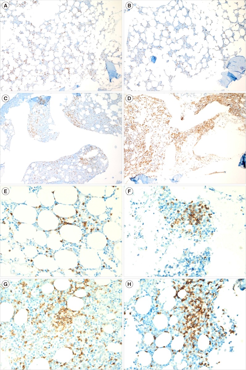
Fig. 1 Representative examples of bone marrow biopsies with various proportions of CD3 positivity in patients with peripheral T-cell lymphoma, not otherwise specified (CD3 immunohistochemical stain, ×100): (A) 10–25%, (B) 26–50%, (C) 51–75%, and (D) 76–100%, and various numbers of CD3-positive cells in a cluster in patients with peripheral T-cell lymphoma, not otherwise specified (CD3 immunohistochemical stain, ×400): (E) <10 cells in a cluster, (F) 21–30 cells, (G) 31–40 cells, and (H) >40 cells.
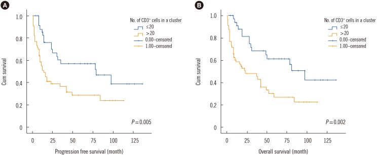
Fig. 2 Kaplan-Meier survival graph showing statistically significant difference in (A) progression free survival and (B) overall survival between >20 and ≤20 CD3+ cells in a cluster.
Reference
-
1. Savage KJ. Update: peripheral T-cell lymphomas. Curr Hematol Malig Rep. 2011; 6:222–230. PMID: 21953415.2. Delmer A, Caulet S, Bryard F, Tabah I, Audouin J, Le Tourneau A, et al. Peripheral T-cell malignant lymphomas. Clinical, morphologic and developmental features in 22 cases [Article in French]. Presse Med. 1990; 19:851–855. PMID: 2140179.3. Chott A, Augustin I, Wrba F, Hanak H, Ohlinger W, Radaszkiewicz T. Peripheral T-cell lymphomas: a clinicopathologic study of 75 cases. Hum Pathol. 1990; 21:1117–1125. PMID: 2227919.4. Gisselbrecht C, Gaulard P, Lepage E, Coiffier B, Brière J, Haioun C, et al. Prognostic significance of T-cell phenotype in aggressive non-Hodgkin's lymphomas. Groupe d'Etudes des Lymphomes de l'Adulte (GELA). Blood. 1998; 92:76–82. PMID: 9639502.5. López-Guillermo A, Cid J, Salar A, López A, Montalbán C, Castrillo JM, et al. Peripheral T-cell lymphomas: initial features, natural history, and prognostic factors in a series of 174 patients diagnosed according to the R.E.A.L. Classification. Ann Oncol. 1998; 9:849–855. PMID: 9789607.6. Dogan A, Morice WG. Bone marrow histopathology in peripheral T-cell lymphomas. Br J Haematol. 2004; 127:140–154. PMID: 15461619.7. Carbone PP, Kaplan HS, Musshoff K, Smithers DW, Tubiana M. Report of the committee on Hodgkin’s disease staging classification. Cancer Res. 1971; 31:1860–1861. PMID: 5121694.8. Oken MM, Creech RH, Tormey DC, Horton J, Davis TE, McFadden ET, et al. Toxicity and response criteria of the Eastern Cooperative Oncology Group. Am J Clin Oncol. 1982; 5:649–655. PMID: 7165009.9. The International Non-Hodgkin's Lymphoma Prognostic Factors Project. A predictive model for aggressive non-Hodgkin's lymphoma. N Engl J Med. 1993; 329:987–994. PMID: 8141877.10. Gallamini A, Stelitano C, Calvi R, Bellei M, Mattei D, Vitolo U, et al. Peripheral T-cell lymphoma unspecified (PTCL-U): a new prognostic model from a retrospective multicentric clinical study. Blood. 2004; 103:2474–2479. PMID: 14645001.11. Falini B, Pileri S, De Solas I, Martelli MF, Mason DY, Delsol G, et al. Peripheral T-cell lymphoma associated with hemophagocytic syndrome. Blood. 1990; 75:434–444. PMID: 2153036.12. Rao SA, Gottesman SR, Nguyen MC, Braverman AS. T cell lymphoma associated with myelofibrosis. Leuk Lymphoma. 2003; 44:715–718. PMID: 12769351.13. Weisenburger DD, Savage KJ, Harris NL, Gascoyne RD, Jaffe ES, MacLennan KA, et al. Peripheral T-cell lymphoma, not otherwise specified: a report of 340 cases from the International Peripheral T-cell Lymphoma Project. Blood. 2011; 117:3402–3408. PMID: 21270441.
- Full Text Links
-
- Actions
-
Cited
- CITED
-
- Close
- Share
-
- Similar articles
-
- A Case of Primary Bone Marrow Lymphoma Secondary to Aplastic Anemia
- A Case of Bone Marrow Involvement of Hepatosplenic gamma delta-Cell Lymphoma
- Peripheral T‑cell lymphoma, NOS in bone marrow and heart
- A Case of Hepatosplenic T-cell Lymphoma Diagnosed by Bone Marrow Examination
- Two Cases of Bone Marrow Permeated Primary Gastric Mucosa-Associated Lymphoid Tissue Lymphoma
