J Korean Ophthalmol Soc.
2010 May;51(5):764-768.
Surgical Treatment of Extensive Conjunctival Melanocytic Nevus Mimicking Conjunctival Melanoma
- Affiliations
-
- 1Department of Ophthalmology, Seoul National University College of Medicine, Seoul Artificial Eye Center, Seoul National University Hospital Clinical Research Institute, Seoul, Korea. eyeminerva@yahoo.co.kr
- 2Department of Ophthalmology, Seoul National University Hospital Boramae Hospital, Seoul, Korea.
- 3Seoul National University Hospital, Health Care System Gangnam Center, Healthcare Research Institute, Seoul, Korea.
Abstract
- PURPOSE
To report a case of diffuse conjunctival melanocytic lesion mimicking conjunctival melanoma and treated by surgical excision and amniotic membrane transplantation.
CASE SUMMARY
A 29-year-old man presented with diffuse pigmented lesion on the bulbar conjunctiva in the right eye, which had been present since birth. Circumferential pigmentation was observed in the perilimbal conjunctiva from 4 to 11 o'clock, and slightly elevated, dark brown-colored lesions with multiple small cysts were noted on the superior, inferior, and temporal bulbar conjunctiva. Incisional biopsy was performed from multiple sites to rule out conjunctival melanoma. Histopathologic examination showed small nevus cells and multiple cysts. Under local anesthesia, temporal conjunctival excision and amniotic membrane transplantation were performed. The surgical pathologist confirmed compound nevus. Four weeks after the surgery, full epithelialization was observed over the amniotic membrane. Several lesions were intentionally left during the surgery, and unnoticeable from the frontal view. The patient was satisfied with the surgical result.
CONCLUSIONS
In extensive conjunctival pigmented lesion, biopsy should always be performed to rule out melanoma. Temporal conjunctival excision rather than whole lesion excision can be a cosmetically good surgical option for a diffuse conjuntival lesion proven as a benign conjunctival nevus.
Keyword
MeSH Terms
Figure
-
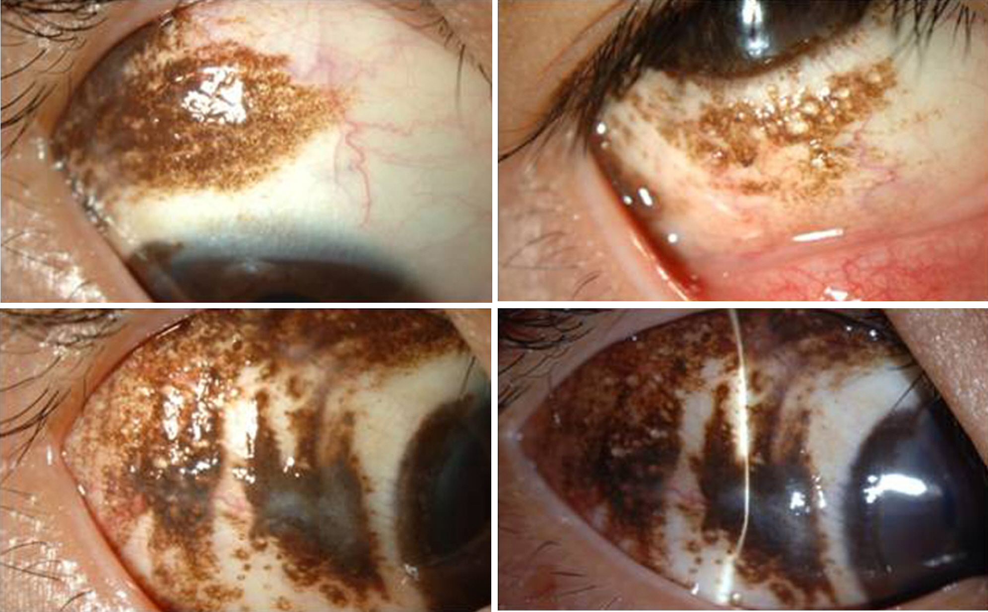
Figure 1. Circumferential pigmentation was observed on the limbus from 4 to 11 o'clock, and slightly elevated, dark brown-colored lesions with multiple small cysts were noted on the superior (Top left), inferior (Top right), and temporal (Bottom left) bulbar conjunctiva. On the slitlamp examination, the lesions were proved to be slightly elevated (Bottom right).

Figure 2. Compared with the preoperative photograph (A) the postoperative photograph (B) showed no noticeable pigmented lesion on the front view.
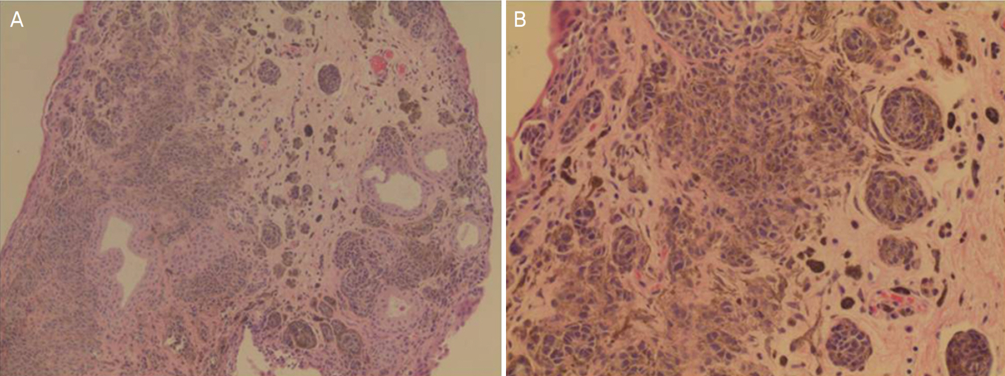
Figure 3. Histopathologic examinations showed that multiple nests of nevus cells and several stromal cysts. (A) ×200, (B) ×400.
Reference
-
References
1. Kurli M, Finger PT. Melanocytic conjunctival tumors. Ophthalmol Clin North Am. 2005; 18:15–24.
Article2. Grossniklaus HE, Green WR, Luckenbach M, Chan CC. Conjunctival lesions in adults: a clinical and histopathological review. Cornea. 1987; 6:78–116.3. Shields CL, Fasiuddin AF, Mashayekhi A, Shields JA. Conjunctival ne-vi: clinical features and natural course in 410 consecutive pateints. Arch Ophthalmol. 2004; 122:167–75.4. Jeoung JW, Kim T, Lee JH, et al. Argon laser ablation of conjunctival nevus. J Korean Ophthalmol Soc. 2004; 45:1989–94.5. Park JJ, Jeong BJ, Seo HD, et al. Treatment of conjunctival nevus with argon laser. J Korean Ophthalmol Soc. 2004; 45:1995–9.6. Tomita M, Goto H, Muramatsu R, Usui M. Treatment of large abdominal nevus by resection and reconstruction using amniotic membrane. Graefes Arch Clin Exp Ophthalmol. 2006; 244:761–4.7. Ash JE. Epibulbar tumors. Am J Ophthalmol. 1950; 33:1203–19.
Article8. Shields CL, Demirci H, Karatza E, et al. Clinical survey of 1643 abdominal and nonmelanocytic conjunctival tumors. Ophthalmology. 2004; 111:1747–54.9. Lee HS, Lew HL, Yun YS, Sim JY. Pigmented spindle cell nevus of the palpebral conjunctiva. J Korean Ophthalmol Soc. 2002; 43:2589–92.10. Kim SY, Lee SB, Yang SW. A case of conjunctival malignant abdominal with extensive corneal displacement. J Korean Ophthalmol Soc. 2005; 46:1235–9.11. Maly A, Epstein D, Meir K, Pe'er J. Histological criteria for grading of atypia in melanocytic conjunctival lesions. Pathology. 2008; 40:676–81.
Article12. Shields CL, Shields JA. Overview of tumors of the conjunctiva and cornea. Foster CS, Azar DT, Dohlman CH, editors. Smolin and Troft's the cornea. 4th ed.Philadelphia: Williams & Wilkins;2004. chap. 40.13. Colby K, Harissi-Dagher M. Tumors of the cornea and conjunctiva. Albert DM, Miller JW, editors. Principles and practice of abdominal. 3rd ed.Philadelphia: Elsevier Inc.;2008. 3:chap. 58.14. Shields JA, Shields CL. Surgical management of conjunctival tumors. Atlas of Eyelid and Conjunctival Tumors. Philadelphia: Lippincott Williams Wilkins;1999. chap. 25.
Article15. Shields CL, Shields JA, Amstrong T. Management of conjunctival and corneal melanoma with surgical excision, amniotic membrane allograft, and topical chemotherapy. Am J Ophthalmol. 2001; 132:576–8.
Article16. Paridaens D, Beekhuis H, van Den Bosch W, et al. Amniotic abdominal transplantation in the management of conjunctival malignant melanoma and primary acquired melanosis with atypia. Br J Ophthalmol. 2001; 85:658–61.17. Tseng SC, Prabhasawat P, Lee SH. Amniotic membrane abdominal for conjunctival surface reconstruction. Am J Ophthalmol. 1997; 124:765–74.
- Full Text Links
-
- Actions
-
Cited
- CITED
-
- Close
- Share
-
- Similar articles
-
- A Case of Giant Conjunctival Nevus Mimicking Malignant Melanoma
- Three Cases of Malignant Melanoma Possibly Arising in a Long Standing Melanocytic Nevus
- A Case of Conjunctival Malignant Melanoma with Extensive Corneal Displacement
- Clinical Manifestations of Extruded Conjunctival Melanocytic Mass
- Malignant Melanoma on Congenital Melanocytic Nevus
