Craniotomy and Membranectomy for Treatment of Organized Chronic Subdural Hematoma
- Affiliations
-
- 1Department of Neurosurgery, Kyungpook National University Hospital, Kyungpook National University School of Medicine, Daegu, Korea. nsdoctor@naver.com
- KMID: 2424326
- DOI: http://doi.org/10.13004/kjnt.2018.14.2.134
Abstract
- We report the case of a patient with organized chronic subdural hematoma (OCSH) that was treated with craniotomy. A 72-year-old man was admitted with a complaint of a drowsy mental status after a generalized tonic-clonic seizure. A brain computed tomography scan acquired at a local hospital revealed a large chronic subdural hematoma (CSDH) in the left frontoparietal lobe. The patient had not experienced head trauma and had been taking clopidogrel due to angina. A neurosurgeon at the local hospital performed single burr hole trephination in the left frontal bone and drained some of the hematoma. Brain magnetic resonance imaging performed upon transfer to our hospital showed a large OCSH with a midline shift to the right side, revealing a low, heterogeneous signal on T2-weighted images (WI) and an isodense signal on T1-WI. We performed craniotomy and membranectomy to achieve adequate decompression and expansion of the brain. Following this, the patient recovered completely. Our findings support that neurosurgeons should consider the possibility of organization of a CSDH when selecting a diagnosis and treatment plan.
Keyword
MeSH Terms
Figure
-
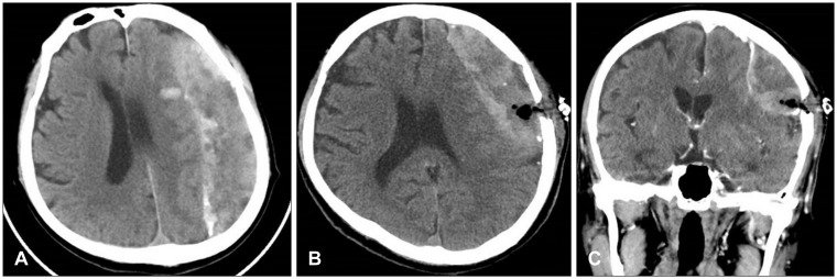
FIGURE 1 Brain computed tomography (CT) scan (A) showed chronic subdural hematoma containing many high density areas in the hematoma and the membrane in the left frontoparietal lobe with a midline shift. After burr hole surgery, brain CT axial (B) and coronal (C) scans demonstrated insufficient drainage of the hematoma through a single burr hole.
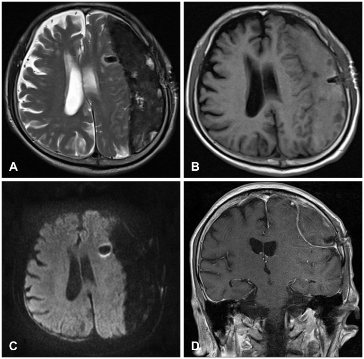
FIGURE 2 Brain magnetic resonance imaging showed a large amount of chronic subdural hematoma with a low, heterogeneous signal on T2-weighted images (WI) (A), an isodense signal on T1-WI (B), and a low, homogeneous signal on diffusion-WI (C). There was no strong enhancement and the membrane surrounding the hematoma was enhanced in a line (D).
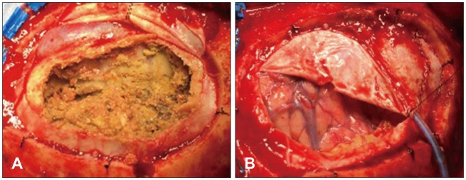
FIGURE 3 Operative findings showed organized chronic subdural hematoma with a thick membrane (A). Note the normal brain cortex after membranectomy (B).
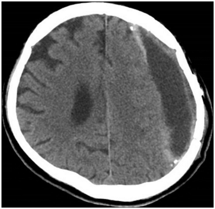
FIGURE 4 Brain computed tomography showed no recurrence of chronic subdural hematoma with subdural fluid collection.
Reference
-
1. Callovini GM, Bolognini A, Callovini G, Gammone V. Primary enlarged craniotomy in organized chronic subdural hematomas. Neurol Med Chir (Tokyo). 2014; 54:349–356. PMID: 24305027.
Article2. Fujioka M, Okuchi K, Miyamoto S, Sakaki T, Tsunoda S, Iwasaki S. Bilateral organized chronic subdural haematomas: high field magnetic resonance images and histological considerations. Acta Neurochir (Wien). 1994; 131:265–269. PMID: 7754833.
Article3. Hellwig D, Heinze S, Riegel T, Benes L. Neuroendoscopic treatment of loculated chronic subdural hematoma. Neurosurg Clin N Am. 2000; 11:525–534. PMID: 10918025.
Article4. Isobe N, Sato H, Murakami T, Kurokawa Y, Seyama G, Oki S. Six cases of organized chronic subdural hematoma. No Shinkei Geka. 2008; 36:1115–1120. PMID: 19086442.5. Kawano N, Endo M, Saito M, Nakayama K, Beppu T. Origin and pathological significance of smooth muscle cells and myofibroblasts in the subdural neomembrane. Neurol Med Chir (Tokyo). 1986; 26:361–368. PMID: 2429215.
Article6. Lee JY, Ebel H, Ernestus RI, Klug N. Various surgical treatments of chronic subdural hematoma and outcome in 172 patients: is membranectomy necessary? Surg Neurol. 2004; 61:523–527. discussion 527-528. PMID: 15165784.
Article7. Oda S, Shimoda M, Hoshikawa K, Shiramizu H, Matsumae M. Organized chronic subdural haematoma with a thick calcified inner membrane successfully treated by surgery: a case report. Tokai J Exp Clin Med. 2010; 35:85–88. PMID: 21319032.8. Rocchi G, Caroli E, Salvati M, Delfini R. Membranectomy in organized chronic subdural hematomas: indications and technical notes. Surg Neurol. 2007; 67:374–380. PMID: 17350406.
Article9. Shrestha P, Pant B, Shrestha P, Rajbhandari P. Organized subdural hematoma with thick membrane in chronic subdural hematoma. JNMA J Nepal Med Assoc. 2012; 52:1–5. PMID: 23279765.
Article10. Suyama T, Ubagai R, Inui T, Tominaga S. Chronic subdural hematoma organized over a short period of time with spontaneous intracranial hypotension: a case report. Brain Nerve. 2012; 64:855–860. PMID: 22764357.
- Full Text Links
-
- Actions
-
Cited
- CITED
-
- Close
- Share
-
- Similar articles
-
- Chronic Subdural Hematoma Treated by Small or Large Craniotomy with Membranectomy as the Initial Treatment
- A Case of Chronic Subdural Hematoma of Infact Developing Communicating Hydrocephalus
- An Organized Chronic Subdural Hematoma with Partial Calcification in a Child
- Clinical Observation, of Chronic Subdural Hematomas (Burr hole, Drainage and Craniotomy, Membranectomy)
- Chronic Subdural Hematoma with Calcification: Case Report
