Muscle Thickness and Echo Intensity of the Abdominal and Lower Extremity Muscles in Stroke Survivors
- Affiliations
-
- 1Graduate School of Medical Rehabilitation, Kobe Gakuin University, Kobe, Japan. monjo@avanzar.co.jp
- 2Faculty of Rehabilitation, Kobe Gakuin University, Kobe, Japan.
- KMID: 2424185
- DOI: http://doi.org/10.3988/jcn.2018.14.4.549
Abstract
- BACKGROUND AND PURPOSE
This study compared the muscle thickness (MT) and echo intensity (EI) of the abdominal, thigh, and lower leg muscles between the paretic and nonparetic sides in chronic stroke survivors.
METHODS
Thirty-two stroke survivors living in the community participated in this study. The MT and EI, which are indicators of muscle mass and intramuscular fat or connective tissue, were assessed in the rectus abdominis, external oblique, internal oblique, transversus abdominis, rectus femoris, vastus intermedius, vastus lateralis, vastus medialis, tibialis anterior, gastrocnemius, and soleus via transverse ultrasound imaging. In addition, a possible indicator of physical activity"”the frequency of going out per week"”was evaluated.
RESULTS
All quadriceps muscles and the tibialis anterior were significantly thinner and the EI values of the vastus intermedius, vastus lateralis, vastus medialis, and soleus were significantly higher in the paretic limb than the nonparetic limb. The MT and EI values of abdominal muscles did not differ significantly between the two sides. The MT values of the paretic rectus femoris, vastus lateralis, and vastus medialis were significantly associated with the frequency of going out after adjusting confounding factors. The MT of the nonparetic vastus lateralis was significantly associated with latency from stroke onset after adjusting confounding factors.
CONCLUSIONS
Our results indicate that quantitative and qualitative changes on the paretic side in stroke survivors were the most robust in the thigh muscles, whereas such changes might not occur in the abdominal muscles.
Keyword
MeSH Terms
Figure
-
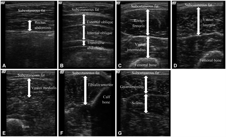
Fig. 1 Muscle ultrasound images and measurement sites and positions. A: Rectus abdominis, at 3 cm lateral to the umbilicus, with the patient supine. B: External oblique, internal oblique, and transverse abdominis, at 2.5 cm anterior to the midaxillary line at the midpoint between the inferior rib and the iliac crest, with the patient supine. C: Rectus femoris and vastus intermedius, at midway between the anterior superior iliac supine and the proximal end of the patella, with the patient supine. D: Vastus lateralis, at midway between the great trochanter and the lateral condyle of the tibia, with the patient supine. E: Vastus medialis, at 30% proximal between the great trochanter and the lateral condyle of the tibia, with the patient supine. F: Tibialis anterior, at 30% proximal between the lateral malleolus of the fibula and the lateral condyle of the tibia, with the patient supine. G: Gastrocnemius, at the medial head of the gastrocnemius at 30% proximal between the lateral malleolus of the fibula and the lateral condyle of the tibia, with the patient sitting. Soleus, at 30% proximal between the lateral malleolus of the fibula and the lateral condyle of the tibia, with the patient sitting.
Cited by 1 articles
-
Changes in Lower Extremity Muscle Quantity and Quality in Patients with Subacute Stroke
Da Hye Kim, Eun Sol Cho, Young Sook Park, Hyun Jung Chang, Jin Gee Park, Jae Yeon Kim, Jeong Hwan Lee
Ann Rehabil Med. 2023;47(6):493-501. doi: 10.5535/arm.23091.
Reference
-
1. Scherbakov N, von Haehling S, Anker SD, Dirnagl U, Doehner W. Stroke induced sarcopenia: muscle wasting and disability after stroke. Int J Cardiol. 2013; 170:89–94. PMID: 24231058.
Article2. Hafer-Macko CE, Ryan AS, Ivey FM, Macko RF. Skeletal muscle changes after hemiparetic stroke and potential beneficial effects of exercise intervention strategies. J Rehabil Res Dev. 2008; 45:261–272. PMID: 18566944.
Article3. English C, McLennan H, Thoirs K, Coates A, Bernhardt J. Loss of skeletal muscle mass after stroke: a systematic review. Int J Stroke. 2010; 5:395–402. PMID: 20854624.
Article4. Ryan AS, Buscemi A, Forrester L, Hafer-Macko CE, Ivey FM. Atrophy and intramuscular fat in specific muscles of the thigh: associated weakness and hyperinsulinemia in stroke survivors. Neurorehabil Neural Repair. 2011; 25:865–872. PMID: 21734070.5. Prado-Medeiros CL, Silva MP, Lessi GC, Alves MZ, Tannus A, Lindquist AR, et al. Muscle atrophy and functional deficits of knee extensors and flexors in people with chronic stroke. Phys Ther. 2012; 92:429–439. PMID: 22135704.
Article6. Goodpaster BH, Carlson CL, Visser M, Kelley DE, Scherzinger A, Harris TB, et al. Attenuation of skeletal muscle and strength in the elderly: the Health ABC Study. J Appl Physiol. 2001; 90:2157–2165. PMID: 11356778.
Article7. Fukumoto Y, Ikezoe T, Yamada Y, Tsukagoshi R, Nakamura M, Mori N, et al. Skeletal muscle quality assessed from echo intensity is associated with muscle strength of middle-aged and elderly persons. Eur J Appl Physiol. 2012; 112:1519–1525. PMID: 21847576.
Article8. Ryan AS, Dobrovolny CL, Smith GV, Silver KH, Macko RF. Hemiparetic muscle atrophy and increased intramuscular fat in stroke patients. Arch Phys Med Rehabil. 2002; 83:1703–1707. PMID: 12474173.
Article9. Ramsay JW, Barrance PJ, Buchanan TS, Higginson JS. Paretic muscle atrophy and non-contractile tissue content in individual muscles of the post-stroke lower extremity. J Biomech. 2011; 44:2741–2746. PMID: 21945568.
Article10. Lee SS, Spear S, Rymer WZ. Quantifying changes in material properties of stroke-impaired muscle. Clin Biomech (Bristol, Avon). 2015; 30:269–275.
Article11. Berenpas F, Martens AM, Weerdesteyn V, Geurts AC, van Alfen N. Bilateral changes in muscle architecture of physically active people with chronic stroke: a quantitative muscle ultrasound study. Clin Neurophysiol. 2017; 128:115–122. PMID: 27888744.
Article12. Pillen S, Tak RO, Zwarts MJ, Lammens MM, Verrijp KN, Arts IM, et al. Skeletal muscle ultrasound: correlation between fibrous tissue and echo intensity. Ultrasound Med Biol. 2009; 35:443–446. PMID: 19081667.
Article13. Bohannon RW, Cassidy D, Walsh S. Trunk muscle strength is impaired multidirectionally after stroke. Clin Rehabil. 1995; 9:47–51.
Article14. Hsieh CL, Sheu CF, Hsueh IP, Wang CH. Trunk control as an early predictor of comprehensive activities of daily living function in stroke patients. Stroke. 2002; 33:2626–2630. PMID: 12411652.
Article15. Dettmann MA, Linder MT, Sepic SB. Relationships among walking performance, postural stability, and functional assessments of the hemiplegic patient. Am J Phys Med. 1987; 66:77–90. PMID: 3578493.16. Duncan PW, Propst M, Nelson SG. Reliability of the Fugl-Meyer assessment of sensorimotor recovery following cerebrovascular accident. Phys Ther. 1983; 63:1606–1610. PMID: 6622535.
Article17. Hebert JJ, Koppenhaver SL, Parent EC, Fritz JM. A systematic review of the reliability of rehabilitative ultrasound imaging for the quantitative assessment of the abdominal and lumbar trunk muscles. Spine. 2009; 34:E848–E856. PMID: 19927091.
Article18. Koppenhaver SL, Hebert JJ, Fritz JM, Parent EC, Teyhen DS, Magel JS. Reliability of rehabilitative ultrasound imaging of the transversus abdominis and lumbar multifidus muscles. Arch Phys Med Rehabil. 2009; 90:87–94. PMID: 19154834.
Article19. Ikezoe T, Nakamura M, Shima H, Asakawa Y, Ichihashi N. Association between walking ability and trunk and lower-limb muscle atrophy in institutionalized elderly women: a longitudinal pilot study. J Physiol Anthropol. 2005; 28:34–31.
Article20. Miyatani M, Kanehisa H, Azuma K, Kuno S, Fukunaga T. Site-related differences in muscle loss with aging. Int J Sport Health Sci. 2003; 1:34–40.
Article21. Abe T, Sakamaki M, Yasuda T, Bemben MG, Kondo M, Kawakami Y, et al. Age-related, site-specific muscle loss in 1507 Japanese men and women aged 20 to 95 years. J Sports Sci Med. 2011; 10:145–150. PMID: 24149307.22. Abe T, Loenneke JP, Thiebaud RS, Fukunaga T. Age-related site-specific muscle wasting of upper and lower extremities and trunk in Japanese men and women. Age (Dordr). 2014; 36:813–821. PMID: 24243442.
Article23. English C, Healy GN, Coates A, Lewis L, Olds T, Bernhardt J. Sitting and activity time in people with stroke. Phys Ther. 2016; 96:193–201. PMID: 26112254.
Article24. Verschuren O, Mead G, Visser-Meily A. Sedentary behaviour and stroke: foundational knowledge is crucial. Transl Stroke Res. 2015; 6:9–12. PMID: 25216691.
Article25. Hachisuka K, Umezu Y, Ogata H. Disuse muscle atrophy of lower limbs in hemiplegic patients. Arch Phys Med Rehabil. 1997; 78:13–18. PMID: 9014951.
Article26. Eng JJ, Lomaglio MJ, Macintyre DL. Muscle torque preservation and physical activity in individuals with stroke. Med Sci Sports Exerc. 2009; 41:1353–1360. PMID: 19516167.
Article27. Billinger SA, Arena R, Bernhardt J, Eng JJ, Franklin BA, Johnson CM, et al. Physical activity and exercise recommendations for stroke survivors: a statement for healthcare professionals from the American Heart Association/American Stroke Association. Stroke. 2014; 45:2532–2553. PMID: 24846875.28. Katano H, Ohno M, Yamada K. Protection by physical activity against deleterious effect of smoking on carotid intima-media thickness in young Japanese. J Stroke Cerebrovasc Dis. 2013; 22:176–183. PMID: 22056218.
Article29. Laurin D, Verreault R, Lindsay J, MacPherson K, Rockwood K. Physical activity and risk of cognitive impairment and dementia in elderly persons. Arch Neurol. 2001; 58:498–504. PMID: 11255456.
Article30. Lytle ME, Vander Bilt J, Pandav RS, Dodge HH, Ganguli M. Exercise level and cognitive decline: the MoVIES project. Alzheimer Dis Assoc Disord. 2004; 18:57–64. PMID: 15249848.31. Laufer Y, Sivan D, Schwarzmann R, Sprecher E. Standing balance and functional recovery of patients with right and left hemiparesis in the early stages of rehabilitation. Neurorehabil Neural Repair. 2003; 17:207–213. PMID: 14677216.
Article32. Patterson KK, Gage WH, Brooks D, Black SE, McIlroy WE. Changes in gait symmetry and velocity after stroke: a cross-sectional study from weeks to years after stroke. Neurorehabil Neural Repair. 2010; 24:783–790. PMID: 20841442.
Article33. Kim HD, You JM, Han N, Eom MJ, Kim JG. Ultrasonographic measurement of transverse abdominis in stroke patients. Ann Rehabil Med. 2014; 38:317–326. PMID: 25024954.
Article
- Full Text Links
-
- Actions
-
Cited
- CITED
-
- Close
- Share
-
- Similar articles
-
- Changes in Muscle Thickness and Echo Intensity in Chronic Stroke Survivors: A 2-Year Longitudinal Study
- The Effect of Lower Extremity Strengthening Exercise Using Sliding Stander on Balance and Spasticity in Chronic Stroke: A Randomized Clinical Trial
- Musculoskeletal Problems in Lower Extremity after Stroke
- Changes in Lower Extremity Muscle Quantity and Quality in Patients with Subacute Stroke
- Ultrasonographic Measurement of Transverse Abdominis in Stroke Patients
