Study on Airway Size in Class III Malocclusion by the Gender
- Affiliations
-
- 1Department of Dental Hygiene, Cheongam College, Korea. smili@hanmail.net
- KMID: 2424166
- DOI: http://doi.org/10.11637/kjpa.2017.30.4.145
Abstract
- The purpose of this study was to investigate the difference of airway between skeletal Class III malocclusion according to gender and to analyze the relationship between airway size and oral maxillofacial position. The total of 40 study subjects was sampled by judgment sampling, who were under 1 degree ANB and who wanted orthognathic surgery. They were taken with lateral cephalometric radiograph and the photos were used for final analysis. The variables were calculated using mean and standard deviation, those were analyzed by independent t-test, Pearson's correlation coefficient and multiple regression analysis. It was located forward than the female that was the measurements of frontal bone, maxilla, mandible, and mentum in the male. The laryngeal pharynx size of the hard and soft tissues was also longer than the female. It was not statistically significant that was the correlation between the variables and the oral pharynx size. However, the width of the laryngeal pharynx increased with male and mandibular protrusion, and it was increased with the position of the anterior of frontal bone, maxilla, and mentum. The variables affecting the airway size are the laryngeal pharynx width of the hard and soft tissue.
MeSH Terms
Figure
-
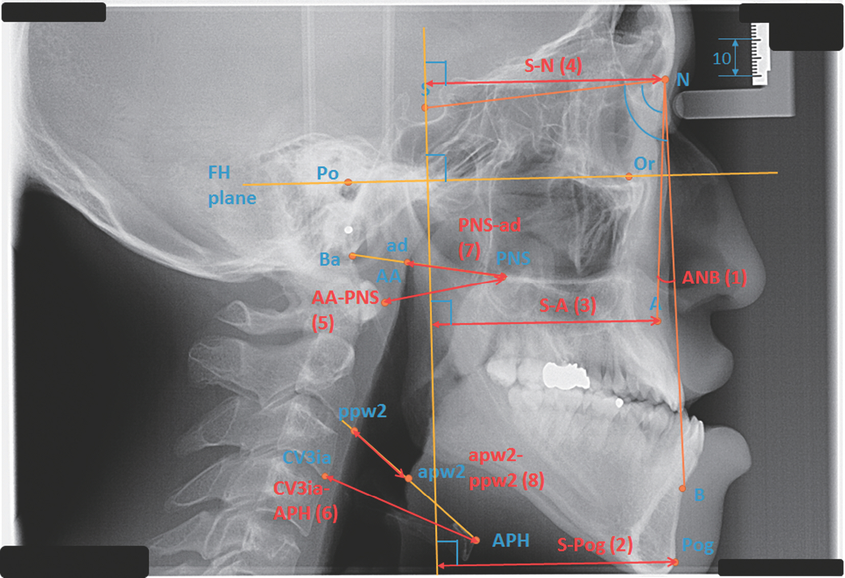
Fig. 1. Craniometric point, plane, angle, and distance. S (Sella turcica, the central point of pituitary gland of sphenoid bone), N (Nasion, the nodal point between sutura of nasal bone and frontal bone and sutura of nasal bone) Or (Orbitale, the most inferior point of orbit), PNS (Posterior nasal spine, the most posterior point of hard palate), A (A point, the most posterior point of curvature lipping between fundus of mandible and alveolar bone), B (B point, the most posterior point of median sutura of mandibular outline), Pog (Pogonion, the most anterior point of median sutura of mandibular outline), Ba (Basion, the most anterior inferior point of greater occipital foramen), Po (Porion, the most superior point of external acoustic meatus), APH (the most anterior point of hyoid bone), AA (the most anterior point of anterior arch of 1st atlas), CV3ia (the most inferior point of 3rd atlas), ad (the nodal point between posterior pharyngeal wall and connecting line from PNS to Ba), apw2 (the nodal point between anterior pharyngeal wall and connecting line from the most anterior point of hyoid bone to the most anterior point of 2nd atlas), ppw2 (the nodal point between posterior pharyngeal wall and connecting line from the most anterior point of hyoid bone to the most anterior posterior point of 2nd atlas).
Reference
-
References
1. Hwang YI, Lee KH, Lee KJ, Kim SC, Cho HJ, Cheon SH, et al. Effect of airway and tongue in facial morphology of prepubertal Class Ⅰ, Ⅱchildren. Korea J Orthod. 2008; 38:74–82.2. Park SH, Yu HS. A comparison study of the effects of hy-pertrophied adenoid tissue on jaws morphology. Korea J Orthod. 2002; 32:19–31.3. Lee MJ, Kim JG, Yang YM, Baik BJ. Effects of mouth breathing on facial skeletal morphology. J Korean Acad Pediatr Dent. 2012; 39:339–47.
Article4. Jung HL, Cha KS, Chung DW. A study on the correlation between airway space and facial morphology in Class III malocclusion children with nasal obstruction. Korean J Orthod. 2007; 37:192–203.5. Lee YS, Kim JC. A cephalometric study on the airway size according to the types of the malocclusion. Korean J Orthod. 1995; 25:19–29.6. Faul F, Erdfelder E, Lang AG, Buchner A. G∗Power 3: A flexible statistical power analysis program for the social, behavioral, and biomedical science. Behav Res Methods. 2007; 39:175–91.7. Cohen J. Statistical power analysis for behavioral sciences. 2nd ed.Hillsdale: Elsevier;2006. p. 553–8.8. Son WS, Choi YS. Evaluation of hyoid bone position and airway size in Class III malocclusion. Korean J Orthod. 1996; 26:247–54.9. Broadbent BH. A new X-ray technique and its application to orthodontia. Angle Orthod. 1931; 1:45–66.10. King EW. A roentgenographic study of pharyngeal growth. Angle Orthod. 1952; 22:23–37.11. Lowe A, Fleetham JA. Two- and three-dimensional analyses of tongue, airway, and soft palate size. In: Norton ML, Brown ACD (Eds.) Atlas of the Difficult Airway. Mosby Year Book, St. Louis;. 2006. 74–82.12. Pae EK, Lowe AA, Sasaki K, Price C, Tsuchiya M, Fleetham JA. A cephalometric and electromyographic study of upper airway structures in the upright and supine positions. Am J Orthod. 1994; 106:52–9.
Article13. Lyberg T, Krogstad O, Djupesland G. Cephalometric analysis in patients with obstructive sleep apnoea syndrome.:II. Soft tissue morphology. J Laryngol Otol. 1989; 103:293–7.14. Ryan CF, Dickson RI, Lowe AA, Blokmanis A, Fleetham JA. Upper airway measurements predict response to uvu-lopalatopharyngoplasty in obstructive sleep apnea. Laryngoscope. 1990; 100:248–53.
Article15. Maltais F, Carrier G, Cormier Y, Sériès F. Cephalometric measurements in snorers, non-snorers, and patients with sleep apnoea. Thoeax. 1991; 46:419–23.
Article16. Brodie AG. On the growth pattern of the human head. From the third month to the eighth year of life. Am J Anatomy. 1941; 68:209–62.
Article17. Subtely JD. The significance of adenoid tissue in othodon-tia. Angle Orthod. 1954; 24:59–69.18. Handelman CS, Osborne G. Growth of the nasopharynx and adenoid development one to eighteen years. Angle Orthod. 1976; 46:243–59.19. Vig PS, Hall DJ. The inadequacy of cephalometric radiographs for airway assessment. Am J Orthod. 1980; 77:230–2.
Article20. Sora FA, Graber TM, Muller TP. Post pharyngeal lymphoid tissue in Angle Class I and Class II malocclusions. Am J Orthod. 1982; 81:299–309.21. Mergen DC, Jacobs RM. The size of nasopharynx associated with normal occlusion and Class II malocclusion. Angle Orthod. 1970; 40:342–6.22. McNamara JR JA. Influence of respiratory pattern on craniofacial growth. Angle Orthod. 1981; 51:269–300.
- Full Text Links
-
- Actions
-
Cited
- CITED
-
- Close
- Share
-
- Similar articles
-
- Evaluation of hyoid bone position and airway size in Class III malocclusion
- A comparative study of mandibular tooth development between Angle Class I malocclusion group and Angle Class III malocclusion group
- Clinical consideration of Angle's classification Class III malocclusion
- A study on positional change of the hyoid bone before and after activator therapy in angle's Class III malocclusion patients
- A cephalometric study on the airway size according to the types of the malocclusion
