J Korean Fract Soc.
2017 Jul;30(3):131-136. 10.12671/jkfs.2017.30.3.131.
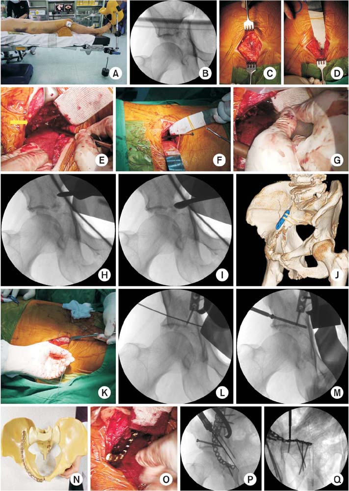
Reduction Technique of Dome Impaction Using the Modified Stoppa Approach: A Technical Note
- Affiliations
-
- 1Department of Orthopedic Surgery, Haeundae Paik Hospital, Inje University College of Medicine, Busan, Korea. bakpaker@hanmail.net
- KMID: 2424121
- DOI: http://doi.org/10.12671/jkfs.2017.30.3.131
Abstract
- In elderly acetabular fractures, central dislocation of the femoral head and impacted superior dome of the acetabulum is common. Unreduced dome impaction can lead to degenerative arthritis and results in poor results. Herein, we present a case of operative reduction and fixation performed via the modified Stoppa approach in acetabular fracture with superior dome impaction.
Figure
Reference
-
1. Anglen JO, Burd TA, Hendricks KJ, Harrison P. The “Gull Sign”: a harbinger of failure for internal fixation of geriatric acetabular fractures. J Orthop Trauma. 2003; 17:625–634.2. Kim JW, Herbert B, Hao J, Min W, Ziran BH, Mauffrey C. Acetabular fractures in elderly patients: a comparative study of low-energy versus high-energy injuries. Int Orthop. 2015; 39:1175–1179.
Article3. Collinge CA, Lebus GF. Techniques for reduction of the quadrilateral surface and dome impaction when using the anterior intrapelvic (modified Stoppa) approach. J Orthop Trauma. 2015; 29:Suppl 2. S20–S24.
Article4. Casstevens C, Archdeacon MT, d'Heurle A, Finnan R. Intrapelvic reduction and buttress screw stabilization of dome impaction of the acetabulum: a technical trick. J Orthop Trauma. 2014; 28:e133–e137.5. Laflamme GY, Hebert-Davies J. Direct reduction technique for superomedial dome impaction in geriatric acetabular fractures. J Orthop Trauma. 2014; 28:e39–e43.
Article6. Garner MR, Sagi HC. Isolated anterior intrapelvic approach for operative reduction and fixation of an anterior column acetabulum fracture with superior impaction. J Orthop Trauma [Internet]. 2016. cited 2016 Jul 12. Available from: http://journals.lww.com/jorthotrauma/Documents/Sept-2016-JOT7994.pdf.7. Kim JW, Kim YC. Modified Stoppa approach in acetabular fractures. J Korean Fract Soc. 2014; 27:274–280.
Article8. Jung GH, Lee Y, Kim JW, Kim JW. Computational analysis of the safe zone for the antegrade lag screw in posterior column fixation with the anterior approach in acetabular fracture: a cadaveric study. Injury. 2017; 48:608–614.
Article9. Moon SW, Kim JW. Usefulness of intraoperative three-dimensional imaging in fracture surgery: a prospective study. J Orthop Sci. 2014; 19:125–131.
Article10. Gary JL, Paryavi E, Gibbons SD, et al. Effect of surgical treatment on mortality after acetabular fracture in the elderly: a multicenter study of 454 patients. J Orthop Trauma. 2015; 29:202–208.
Article11. Jouffroy P; Bone and Joint Trauma Study Group (GETRAUM). Indications and technical challenges of total hip arthroplasty in the elderly after acetabular fracture. Orthop Traumatol Surg Res. 2014; 100:193–197.
Article12. Tosounidis TH, Stengel D, Giannoudis PV. Anteromedial dome impaction in acetabular fractures: issues and controversies. Injury. 2016; 47:1605–1607.
Article13. Sagi HC, Afsari A, Dziadosz D. The anterior intra-pelvic (modified rives-stoppa) approach for fixation of acetabular fractures. J Orthop Trauma. 2010; 24:263–270.
Article14. Scolaro JA, Routt ML Jr. Reduction of osteoarticular acetabular dome impaction through an independent iliac cortical window. Injury. 2013; 44:1959–1964.
Article15. Ma K, Luan F, Wang X, et al. Randomized, controlled trial of the modified Stoppa versus the ilioinguinal approach for acetabular fractures. Orthopedics. 2013; 36:e1307–e1315.
Article16. Hammad AS, El-Khadrawe TA. Accuracy of reduction and early clinical outcome in acetabular fractures treated by the standard ilio-inguinal versus the Stoppa/iliac approaches. Injury. 2015; 46:320–326.
Article17. Kim JW, Shon HC, Park JH. Injury of the obturator nerve in the modified Stoppa approach for acetabular fractures. Orthop Traumatol Surg Res. 2017; DOI: 10.1016/j.otsr.2017.03.005. [epub].
Article18. Zhuang Y, Lei JL, Wei X, Lu DG, Zhang K. Surgical treatment of acetabulum top compression fracture with sea gull sign. Orthop Surg. 2015; 7:146–154.
Article
- Full Text Links
- Actions
-
Cited
- CITED
-
- Close
- Share
- Similar articles
-
- Modified Stoppa Approach in Acetabular Fractures
- Modified Stoppa Approach for Surgical Treatment of Acetabular Fracture
- Adhesion of External Iliac Vessels Found in a Modified Stoppa Approach to Acetabular Fracture in a Patient with a History of Previous Abdominal Surgery
- Perioperative complications of the modified Stoppa approach for the treatment of pelvic bone fractures: a single-institution review of 48 cases
- Direct Brachial Approach for Acute Basilar Artery Occlusion: Technical Note and Preliminary Clinical Experience


