Endovascular Treatment of Idiopathic Intracranial Hypertension with Stenting of the Transverse Sinus Stenosis
- Affiliations
-
- 1Department of Neuroendovascular Therapy Center, Aichi Medical University, Nagakute, Japan. miyachi.shigeru.752@mail.aichi-med-u.ac.jp
- 2Department of Neurosurgery and Endovascular Therapy, Osaka Medical College, Takatsuki, Japan.
- 3Department of Neurosurgery, Ohnishi Neurosurgical Hospital, Akashi, Japan.
- KMID: 2424065
- DOI: http://doi.org/10.5469/neuroint.2018.00990
Abstract
- For many years, the pathophysiology of idiopathic intracranial hypertension (IIH) was interpreted as "secondary intracranial hypertension," and IIH was considered to be caused by brain edema due to obstructive sleep apnea. Another theory proposed cerebrospinal fluid (CSF) absorption impairment due to excessive medication with vitamin A derivatives. Other reports pointed out the importance of obesity, which may cause an impairment of intracranial venous drainage due to elevated right atrial pressure. Patients with medically refractory IIH have traditionally undergone a CSF diversion. Venous outlet impairment on IIH has recently been reported as a causative or contributory cause, and thus focused venoplasty of the stenotic sinus with a stent has emerged as a new treatment strategy. We report the cases of two patients who presented with headache and papilledema with IIH. They successfully underwent stent placement at the stenosis of the transverse sinus and experienced complete resolution of symptoms.
MeSH Terms
Figure
-
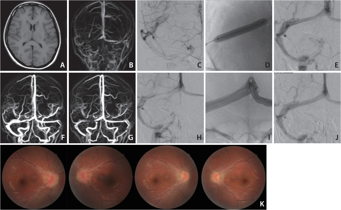
Fig. 1. MRI T1 image shows almost normal findings without hydrocephalus of brain swelling (A). MR venogram demonstrates stenosis of the right TS and occlusion of the left sigmoid sinus (B). A right internal cerebral angiogram shows stenosis of the proximal side of the TS (C). After balloon sinusplasty (D), the TS is well dilated (E). An MR venogram taken 1 week later shows the patency of the TS despite remaining mild stenosis (F). The MR venogram taken after the recurrence of symptoms (POD 42) shows restenosis of the TS (G), and a cerebral angiogram showed recurrence of the TS stenosis as well (H). Postoperative angiogram after the deployment of the stent (I) shows the normalized TS (J). The photo of ocular fundus reveals marked improvement of papilledema. Preoperative image (upper) and at the 4-month follow-up (lower) (K). MRI, magnetic resonance imaging; TS, transverse sinus; POD, postoperative day
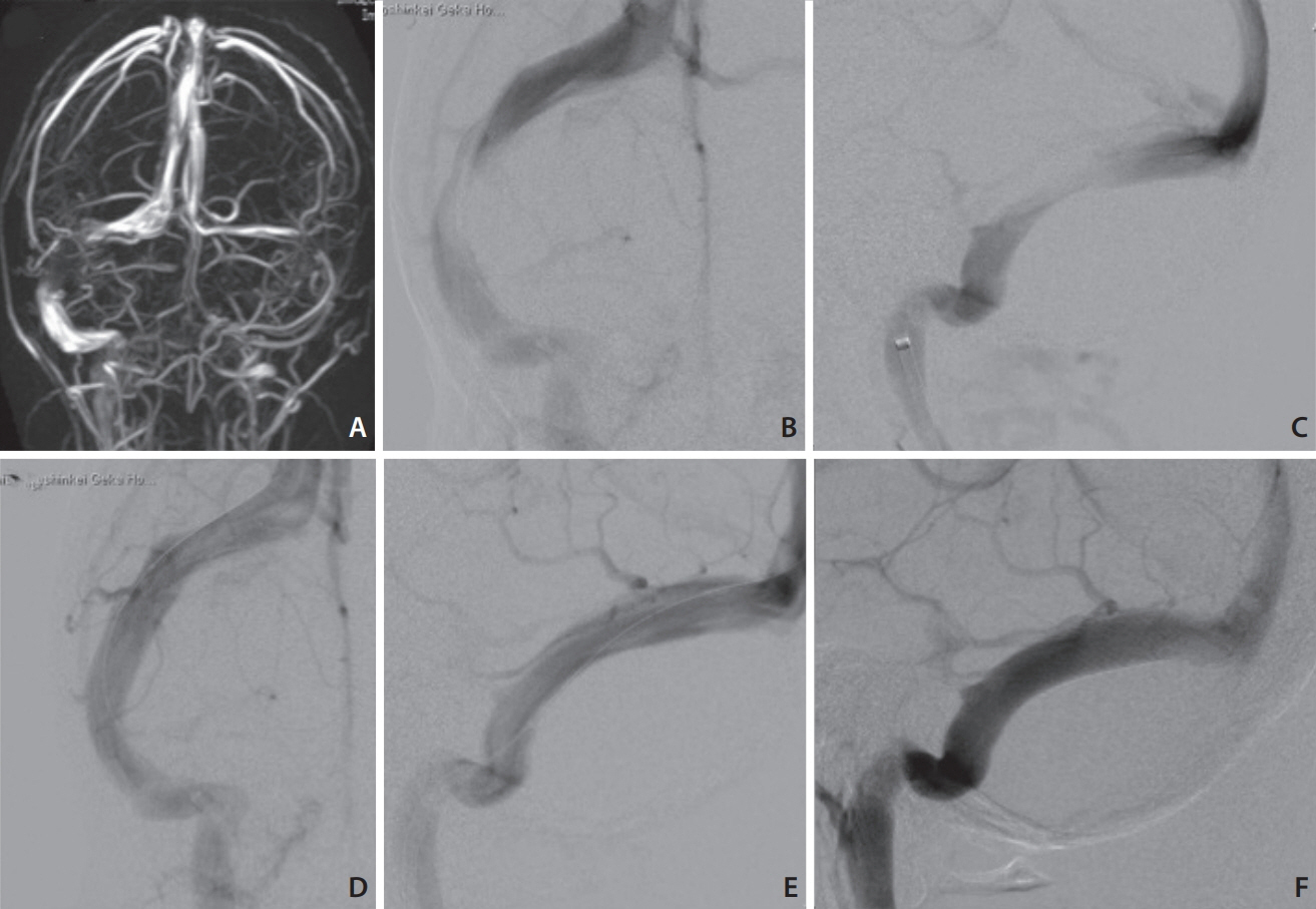
Fig. 2. MR venogram shows stenosis of the mid-portion of the right TS and left internal jugular vein (A). Right internal cerebral angiogram demonstrates severe stenosis of the TS (B: antero-posterior [A-P] view, C: lateral view). Angiogram after stenting shows fully dilated TS (D: A-P view, E: lateral view). Follow-up angiogram (lateral view) 1 month later shows no recurrence (F). MR, magnetic resonance; TS, transverse sinus; A-P, antero-posterior.
Cited by 2 articles
-
Intraluminal anatomy of the transverse sinus: implications for endovascular therapy
Juan J. Altafulla, Joshua Prickett, Joe Iwanaga, Aaron S. Dumont, R. Shane Tubbs
Anat Cell Biol. 2020;53(4):393-397. doi: 10.5115/acb.20.041.Idiopathic Intracranial Hypertension in Asians: A New Perspective and the Need for Scrutiny
Ki Baek Lee, Soo Jeong, Deok Hee Lee
Neurointervention. 2022;17(1):65-66. doi: 10.5469/neuroint.2021.00507.
Reference
-
References
1. Puffer RC, Mustafa W, Lanzino G. Venous sinus stenting for idiopathic intracranial hypertension; a review of the literature. J Neurointervent Surg. 2013; 5:483–486.2. Friedman DI, Jacobson DM. Diagnostic criteria for idiopathic intracranial hypertension. Neurology. 2002; 59:1492–1495.
Article3. Ahmed RM, Wilkinson M, Parker GD, Thurtell MJ, Macdonald J, McCluskey PJ, et al. Transverse sinus stenting for idiopathic intracranial hypertension: a review of 52 patients and of model predictions. AJNR Am J Neuroradiol. 2011; 32:1408–1414.
Article4. Farb RI, Vanek I, Scott JN, Mikulis DJ, Willinsky RA, Tomlinson G, et al. Idiopathic intracranial hypertension: the prevalence and morphology of sinovenous stenosis. Neurology. 2003; 60:1418–1424.
Article5. Higgins JN, Gillard JH, Owler BK, Harkness K, Pickard JD. MR venography in idiopathic intracranial hypertension: unappreciated and misunderstood. J Neurol Neurosurg Psychiatry. 2004; 75:621–625.
Article6. Bussière M, Falero R, Nicolle D, Proulx A, Patel V, Pelz D. Unilateral transverse sinus stenting of patients with idiopathic intracranial hypertension. AJNR Am J Neuroradiol. 2010; 31:645–650.
Article7. King JO, Mitchell PJ, Thomson KR, Tress BM. Manometry combined with cervical puncture in idiopathic intracranial hypertension. Neurology. 2002; 58:26–30.
Article8. Rohr A, Dörner L, Stingele R, Buhl R, Alfke K, Jansen O. Reversibility of venous sinus obstruction in idiopathic intracranial hypertension. AJNR Am J Neuroradiol. 2007; 28:656–659.9. Higgins JN, Owler BK, Cousins C, Pickard JD. Venous sinus stenting for refractory benign intracranial hypertension. Lancet. 2002; 359:228–230.
Article10. Rajpal S, Niemann DB, Turk AS. Transverse venous sinus stent placement as treatment for benign intracranial hypertension in a young male: case report and review of the literature. J Neurosurg. 2005; 102(3 suppl):342–346.11. Binder DK, Horton JC, Lawton MT, McDermott MW. Idiopathic intracranial hypertension. Neurosurgery. 2004; 54:538–551. ; discussion 551-552.
Article12. Acheson JF. Idiopathic intracranial hypertension and visual function. Br Med Bull. 2006; 79–80. 233–240.
Article13. Warner JE, Larson AJ, Bhosale P, Digre KB, Henley C, Alder SC, et al. Retinol-binding protein and retinol analysis in cerebrospinal fluid and serum of patients with and without idiopathic intracranial hypertension. J Neuroophthalmol. 2007; 27:258–262.
Article14. Karahalios DG, Rekate HL, Khayata MH, Apostolides PJ. Elevated intracranial venous pressure as a universal mechanism in pseudotumor cerebri of varying etiologies. Neurology. 1996; 46:198–202.
Article15. Kim TW, Choung HK, Khwarg SI, Hwang JM, Yang HJ. Obesity may not be a risk factor for idiopathic intracranial hypertension in Asians. Eur J Neurol. 2008; 15:876–879.
Article16. Banta JT, Farris BK. Pseudotumor cerebri and optic nerve sheath decompression. Ophthalmology. 2000; 107:1907–1912.
Article17. Satti SE, Leishangthem L, Chaudry MI. Meta-analysis of CSF diversion procedures and dural venous sinus stenting in the setting of medically refractory idiopathic intracranial hypertension. AJNR Am J Neuroradiol. 2015; 36:1899–1904.
Article18. McGirt MJ, Woodworth G, Thomas G, Miller N, Williams M, Rigamonti D. Cerebrospinal fluid shunt placement for pseudotumor cerebri-associated intractable headache: predictors of treatment response and an analysis of long-term outcomes. J Neurosurg. 2004; 101:627–632.
Article19. Kumpe DA, Bennett JL, Seinfeld J, Pelak VS, Chawla A, Tierney M. Dural sinus stent placement for idiopathic intracranial hypertension. J Neurosurg. 2012; 116:538–548.
Article20. Donnet A, Metellus P, Levrier O, Mekkaoui C, Fuentes S, Dufour H, et al. Endovascular treatment of idiopathic intracranial hypertension: clinical and radiologic outcome of 10 consecutive patients. Neurology. 2008; 70:641–647.
Article21. Fields JD, Javedani PP, Falardeau J, Nesbit GM, Dogan A, Helseth EK, et al. Dural venous sinus angioplasty and stenting for the treatment of idiopathic intracranial hypertension. J Neurointerv Surg. 2013; 5:62–68.
Article22. Higgins JN, Cousins C, Owler BK, Sarkies N, Pickard JD. Idiopathic intracranial hypertension: 12 cases treated by venous sinus stenting. J Neurol Neurosurg Psychiatry. 2003; 74:1662–1666.
Article23. Radvany MG, Solomon D, Nijjar S, Subramanian PS, Miller NR, Rigamonti D, et al. Visual and neurological outcomes following endovascular stenting for pseudotumor cerebri associated with transverse sinus stenosis. J Neuroophthalmol. 2013; 33:117–122.
Article24. Stevens SA, Previte M, Lakin WD, Thakore NJ, Penar PL, Hamschin B. Idiopathic intracranial hypertension and transverse sinus stenosis: a modelling study. Math Med Biol. 2007; 24:85–109.
Article25. Teleb MS, Cziep ME, Lazzaro MA, Gheith A, Asif K, Remler B, et al. Idiopathic intracranial hypertension. A systematic analysis of transverse sinus stenting. Interv Neurol. 2013; 2:132–143.
Article
- Full Text Links
-
- Actions
-
Cited
- CITED
-
- Close
- Share
-
- Similar articles
-
- Effect of Transverse Sinus Stenting on Diffuse Leukoencephalopathy with Idiopathic Intracranial Hypertension
- Endovascular Treatment of Idiopathic Intracranial Hypertension: A Case Report
- Angioplasty, Stenting and Other Potential Treatments of Atherosclerotic Stenosis of the Intracranial Arteries: Past, Present and Future
- Guideline for Intracranial Stenting of Symptomatic Intracranial Artery Stenosis: Preliminary Report
- A Case of Intracranial Hypertension with Hypoglycorrhachia Caused by Bilateral Transverse Sinus Stenoses
