In Vitro Evaluation of Fusiform-Shaped Stents for Wide-Neck Intracranial Aneurysm Treatment
- Affiliations
-
- 1Departments of Radiology and Research Institute of Radiology, Asan Medical Center, University of Ulsan College of Medicine, Seoul, Korea. dhlee@amc.seoul.kr
- 2Department of Radiology, The First Affiliated Hospital of Nanjing Medical University, Nanjing, China.
- 3Department of Medical Biotechnology, Dongguk University, Goyang, Korea.
- 4Department of Neurosurgery, Asan Medical Center, University of Ulsan College of Medicine, Seoul, Korea.
- 5Angiovention, Goyang, Korea.
- KMID: 2424060
- DOI: http://doi.org/10.5469/neuroint.2018.00976
Abstract
- PURPOSE
Wide-neck aneurysms (WNAs) associated with a dilated parent artery (PA) are not uncommon morphological abnormalities and usually cause inappropriate wall apposition and incomplete neck coverage of a tubular stent in stent-assisted coiling of aneurysms. We aimed to introduce a fusiform-shaped stent (FSS) and test its effectiveness in treating intracranial WNAs associated with a dilated PA using a three-dimensional (3D) model.
MATERIALS AND METHODS
Two FSS types were designed with the middle one-third segment dilated by 10% (FSS10) and 20% (FSS20) and were compared with the tubular-shaped stent (TSS). A patient-specific 3D WNA model was prototyped and produced, and in vitro stent placement was performed. Angiographic images of the three stent types were analyzed and compared using predetermined parameters.
RESULTS
The stent lumens were significantly larger in FSS10 and FSS20 than in TSS in the middle segments (P=0.046), particularly FSS20 (P=0.018). The non-covered area at the ostium tended to be smaller in FSS10 and FSS20 than in TSS, but the difference was not significant (P>0.05). The stent length was significantly longer in FSS10 and FSS20 than in TSS. The stent cell size was significantly larger in FSS than in TSS.
CONCLUSION
Better vessel wall apposition and aneurysmal neck coverage was observed for FSS than for TSS. No significant difference was observed between FSS10 and FSS20.
Keyword
MeSH Terms
Figure
-

Fig. 1. Stent-assisted WNA coiling and concept design. (A-C) Right internal carotid artery (ICA) ophthalmic segment WNA with PA dilation (aneurysm neck diameter 5.3 mm, proximal PA diameter 4.2 mm, distal PA diameter 2.9 mm, PA diameter at aneurysm neck 5.0 mm) underwent stent-assisted coiling (LVIS blue, 4.5×18 mm). Final three-dimensional angiography showed several loops of coil protruding into the gap between the stent and vessel wall (arrow in B), and the irregular residual space between the stent, coils, and vessel wall (curved arrow in B, stent was visible on C). (D) Illustrations of a TSS and a FSS for coiling of WNAs with a dilated PA. The FSS shows better wall apposition and aneurysmal neck coverage. (E) Prototypes of the TSS and the FSS with middle segment dilated by 10% and 20%. WNA, wide-neck aneurysm; PA, parent artery; TSS, tubular-shaped stent; FSS, fusiform-shaped stent.

Fig. 2. Centerline distance and stent expansion. (A, B) Three-dimensional (3D) volume-rendering image of the aneurysm and the 3D prototype of silicone aneurysm model. (C) Reconstructed 3D image showing the placed stent in the silicone model on the Leonardo workstation. A centerline was drawn along the stented segment of parent artery to measure the length of the stent (purple line). The position of 10 different cross sections perpendicular to the centerline at the aneurysm neck portion was shown, which was used for stent lumen area measurement. (D-L) Representative images (slices 3, 5, and 7 marked in image C) of each type of stent were compared and used for measuring the lumen area of the stents (cut-surface area). Note the gap between the TSS stent and the vessel wall (arrowheads in D), and the better wall apposition evolution from the TSS to FSS20 (arrow in E, H, and K). SL, slices; TSS, tubular-shaped stent; FSS, fusiform-shaped stent.
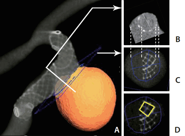
Fig. 3. Stent coverage at the aneurysm neck and cell area at the convex side. (A) Reconstructed 3D image showing the placed stent in the silicone model. (B) A cross section perpendicular to the centerline (corresponding to the white line shown in A). (C) A section at the aneurysm neck to measure the neck coverage of the stent (the blue cutting plane shown in A). The dashed lines between (B) and (C) showing the corresponding points in two planes. On image (C), the stent-covered area and non-covered area of the aneurysm neck can be measured. (D) Measuring the four-cell size at the convex side of the stent.
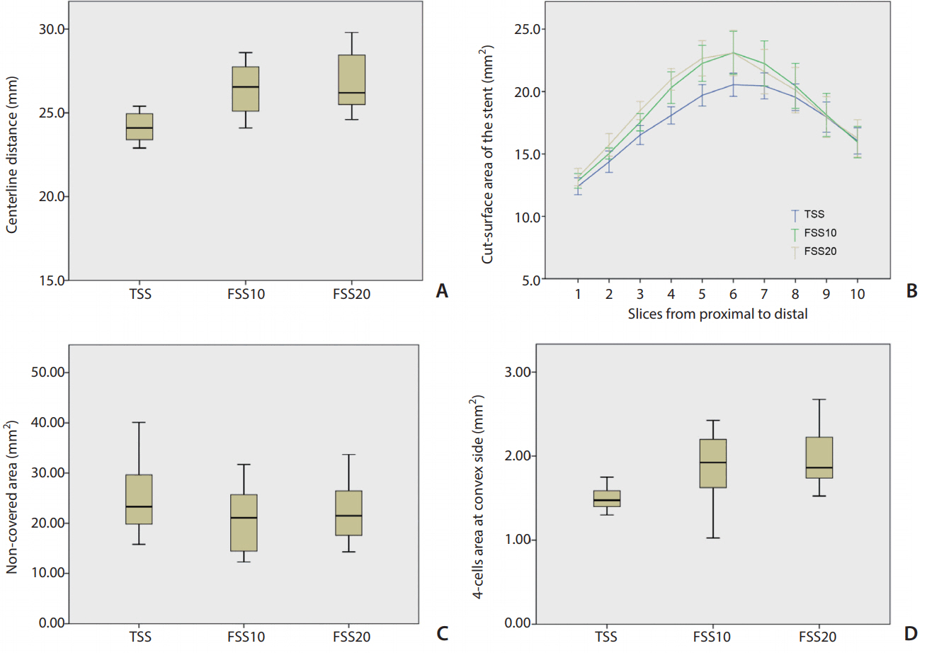
Fig. 4. Analytical results. (A) Comparison of the centerline distances of the stents showing that they are longer for FSS10 and FSS20 than for TSS. (B) Comparison of the stent lumen areas of the 10 sections along the stent among the three types of stents. FSS10 and FSS20 have significantly larger lumen areas than that of the TSS (P=0.024 and P=0.018, respectively). (C) Comparison of the non-covered area sizes at the aneurysmal neck (measured in Fig. 3C) showing no significant difference. (D) Comparison of the four-cell areas at the convex side showing better expansion for FSS10 and FSS20. FSS, fusiform-shaped stent; FSS10, FSS with middle segment dilated by 10%; FSS20, FSS with middle segment dilated by 20%; TSS, tubular-shaped stent.
Reference
-
1. Molyneux AJ, Kerr RS, Yu LM, Clarke M, Sneade M, Yarnold JA, et al. International subarachnoid aneurysm trial (ISAT) of neurosurgical clipping versus endovascular coiling in 2143 patients with ruptured intracranial aneurysms: a randomised comparison of effects on survival, dependency, seizures, rebleeding, subgroups, and aneurysm occlusion. Lancet. 2005; 366:809–817.
Article2. Jeong HW, Seo JH, Kim ST, Jung CK, Suh SI. Clinical practice guideline for the management of intracranial aneurysms. Neurointervention. 2014; 9:63–71.
Article3. Yu M, Liu F, Jiang S, Nie B. Stent-assisted coiling for the treatment of ruptured micro-intracranial wide-necked aneurysms. Interv Neuroradiol. 2015; 21:40–43.
Article4. Fiorella D, Albuquerque FC, Han P, McDougall CG. Preliminary experience using the Neuroform stent for the treatment of cerebral aneurysms. Neurosurgery. 2004; 54:6–16. discussion 16-17.
Article5. Benitez RP, Silva MT, Klem J, Veznedaroglu E, Rosenwasser RH. Endovascular occlusion of wide-necked aneurysms with a new intracranial microstent (Neuroform) and detachable coils. Neurosurgery. 2004; 54:1359–1367. discussion 1368.
Article6. Krischek O, Miloslavski E, Fischer S, Shrivastava S, Henkes H. A comparison of functional and physical properties of self-expanding intracranial stents [Neuroform3, Wingspan, Solitaire, Leo+, Enterprise]. Minim Invasive Neurosurg. 2011; 54:21–28.
Article7. Cho SH, Jo WI, Jo YE, Yang KH, Park JC, Lee DH. Bench-top comparison of physical properties of 4 commercially-available self-expanding intracranial stents. Neurointervention. 2017; 12:31–39.
Article8. Heller RS, Miele WR, Do-Dai DD, Malek AM. Crescent sign on magnetic resonance angiography revealing incomplete stent apposition: correlation with diffusion-weighted changes in stent-mediated coil embolization of aneurysms. J Neurosurg. 2011; 115:624–632.
Article9. Lee JW, Woo JM, Lim OK, Jo Y, Kim JK, Kim ES, et al. Enlarged parent artery lumen at aneurysmal-neck segment in widenecked distal internal carotid artery aneurysms. Neurointervention. 2015; 10:82–88.
Article10. De Bock S, Iannaccone F, De Santis G, De Beule M, Mortier P, Verhegghe B, et al. Our capricious vessels: the influence of stent design and vessel geometry on the mechanics of intracranial aneurysm stent deployment. J Biomech. 2012; 45:1353–1359.
Article11. Lee DH, Hwang SM, Lim OK, Kim JK. In vitro observation of air bubbles during delivery of various detachable aneurysm embolization coils. Korean J Radiol. 2012; 13:412–416.12. Herweh C, Nagel S, Pfaff J, Ulfert C, Wolf M, Bendszus M, et al. First experiences with the new Enterprise2® Stent. Clin Neuroradiol. 2018; 28:201–207.
Article13. Zhang X, Zhong J, Gao H, Xu F, Bambakidis NC. Endovascular treatment of intracranial aneurysms with the LVIS device: a systematic review. J Neurointerv Surg. 2017; 9:553–557.
Article14. Darflinger RJ, Chao K. Using the Barrel technique with the LVIS Jr (low-profile visualized intraluminal support) stent to treat a wide neck MCA bifurcation aneurysm. J Vasc Interv Neurol. 2015; 8:25–27.15. Muhl-Benninghaus R, Simgen A, Reith W, Yilmaz U. The Barrel stent: new treatment option for stent-assisted coiling of widenecked bifurcation aneurysms-results of a single-center study. J Neurointerv Surg. 2017; 9:1219–1222.
- Full Text Links
-
- Actions
-
Cited
- CITED
-
- Close
- Share
-
- Similar articles
-
- Stent-Assisted Coil Trapping in a Manual Internal Carotid Artery Compression Test for the Treatment of a Fusiform Dissecting Aneurysm
- Treatment for Giant Fusiform Aneurysm Located in the Cavernous Segment of the Internal Carotid Artery Using the Pipeline Embolization Device
- Giant Fusiform Aneurysm by Circumferential Wrapping with Sutures-Reinforcement
- Staged Y-shaped Stent Assisted Coil Embolization in a Wide-Neck Basilar Tip Aneurysm: Case Report
- Endovascular Management of the Wide-neck Aneurysms: the Applications of the Coils and Catheter
