MRI Vessel Wall Imaging and Treatment of an Aneurysm at the Atlanto-Axial Segment of an Aberrant Vertebral Artery
- Affiliations
-
- 1Division of Neurosurgery, Department of Surgery, Queen Mary Hospital, The University of Hong Kong, Hong Kong. acotsang@hku.hk
- 2Li Ka Shing Faculty of Medicine, The University of Hong Kong, Hong Kong.
- 3Department of Diagnostic Radiology, Queen Mary Hospital, The University of Hong Kong, Hong Kong.
- KMID: 2424052
- DOI: http://doi.org/10.5469/neuroint.2018.13.1.62
Abstract
- We report a case of unique location of an aneurysm at the atlanto-axial extradural segment of a unilateral aberrant vertebral artery. The MRI vessel wall imaging findings and possible mechanism of aneurysm formation were discussed. A 5 mm extracranial vertebral artery aneurysm located at the interlaminar space between C1 and C2 was diagnosed in a woman presenting with occipital headache. The index vertebral artery ran an aberrant course at the V3 segment, where it entered the dura between C1 and C2 instead of the usual atlanto-occipital space. MR vessel wall imaging showed homogenous wall enhancement of the aneurysm sac. We surmise the anomalous course of the vertebral artery subjected the V3 segment to repeated shearing force secondary to the atlanto-axial rotational neck movement. This led to vessel wall trauma and inflammation, and subsequent aneurysm formation. The aneurysm was successfully treated with endovascular coiling with resolution of symptoms.
Keyword
MeSH Terms
Figure
-
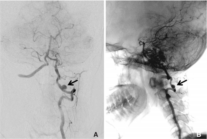
Fig. 1 Catheter angiogram with left vertebral artery injection. (A) Anteroposterior view showing a 5mm saccular aneurysm (arrow) at the extra-dural segment of the vertebral artery. (B) Lateral projection angiogram with fluoroscopy overlay showing the aneurysm (arrow) located at the atlantoaxial level.
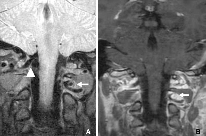
Fig. 2 Coronal MRI showing the aberrant left vertebral artery with the aneurysm located between the left atlas and axis (arrow). (A) PD VISTA and (B) T1-weighted with gadolinium contrast sequence showing circumferential aneurysm wall enhancement. Note the right vertebral artery followed the usual course above the atlas (arrowhead).
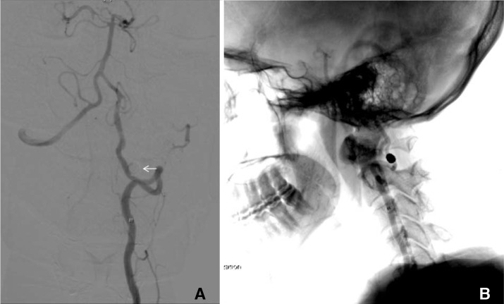
Fig. 3 (A) Control left vertebral artery subtraction angiogram showing its aberrant course, and complete exclusion of aneurysm from the circulation after coil embolization. (B) Lateral projection of the same left vertebral artery angiogram with fluoroscopy overlay. The aneurysm coil mass is located at the interlaminar space between C1 and C2.
Reference
-
1. Shang EK, Fairman RM, Foley PJ, Jackson BM. Endovascular treatment of a symptomatic extracranial vertebral artery aneurysm. J Vasc Surg. 2013; 58:1391–1393. PMID: 23561429.
Article2. Siclari F, Burger IM, Fasel JH, Gailloud P. Developmental anatomy of the distal vertebral artery in relationship to variants of the posterior and lateral spinal arterial systems. AJNR Am J Neuroradiol. 2007; 28:1185–1190. PMID: 17569985.
Article3. Tsang AC, Tsang FC, Lui WM. Bilateral aberrant C1/2 intradural vertebral arteries: a rare cause of cervical myelopathy. ANZ J Surg. 2017; 87:202–203. PMID: 25267558.
Article4. Chung JC, Jung SS, Park KS, Ha HG. Intraoperative vertebral artery angiography to guide c1-2 transarticular screw fixation in a patient with athethoid cerebral palsy. J Korean Neurosurg Soc. 2012; 51:177–181. PMID: 22639719.5. Roche CJ, King SJ, Dangerfield PH, Carty HM. The atlanto-axial joint: physiological range of rotation on MRI and CT. Clin Radiol. 2002; 57:103–108. PMID: 11977941.
Article6. Penning L. Normal movement of the cervical spine. AJR Am J Roentgenol. 1978; 130:317–326. PMID: 414586.7. Edjlali M, Gentric JC, Regent-Rodriguez C, Trystram D, Hassen WB, Lion S, et al. Does aneurysmal wall enhancement on vessel wall MRI help to distinguish stable from unstable intracranial aneurysms? Stroke. 2014; 45:3704–3706. PMID: 25325912.
Article8. Nagahata S, Nagahata M, Obara M, Kondo R, Minagawa N, Sato S, et al. Wall enhancement of the intracranial aneurysms revealed by magnetic resonance vessel wall imaging using three-dimensional turbo spin-echo sequence with motion-sensitized driven-equilibrium: a sign of ruptured aneurysm? Clin Neuroradiol. 2016; 26:277–283. PMID: 25332151.
Article9. Hu P, Yang Q, Wang DD, Guan SC, Zhang HQ. Wall enhancement on high-resolution magnetic resonance imaging may predict an unsteady state of an intracranial saccular aneurysm. Neuroradiology. 2016; 58:979–985. PMID: 27438805.
Article10. Krings T, Mandell DM, Kiehl TR, Geibprasert S, Tymianski M, Alvarez H, et al. Intracranial aneurysms: from vessel wall pathology to therapeutic approach. Nat Rev Neurol. 2011; 7:547–559. PMID: 21931350.
Article11. Qiao Y, Steinman DA, Qin Q, Etesami M, Schär M, Astor BC, et al. Intra-cranial arterial wall imaging using three-dimensional high isotropic resolution black port MRI at 3 Tesla. J Magn Reson Imaging. 2011; 34:22–30. PMID: 21698704.
- Full Text Links
-
- Actions
-
Cited
- CITED
-
- Close
- Share
-
- Similar articles
-
- Author Correction: MRI Vessel Wall Imaging and Treatment of an Aneurysm at the Atlanto-Axial Segment of an Aberrant Vertebral Artery
- High-Resolution Magnetic Resonance Imaging of Intracranial Vertebral Artery Dissecting Aneurysm for Planning of Endovascular Treatment
- Posterior Atlantoaxial Screw-Rod Fixation in a Case of Aberrant Vertebral Artery Course Combined with Bilateral High-Riding Vertebral Artery
- O-arm(R) Imaging System Coupled with Navigation Guided Posterior Atlantoaxial Screw Fixation in the Patient with Ponticulus Posticus, an Anatomical Variation of Atlas
- Total Arch Replacement for Chronic Aortic Aneurysmal Dissection Patient with Aberrant Subclavian Artery
