High-Resolution Magnetic Resonance Imaging of Intracranial Vertebral Artery Dissecting Aneurysm for Planning of Endovascular Treatment
- Affiliations
-
- 1Department of Neurosurgery, Busan Paik Hospital, Inje University College of Medicine, Busan, Korea. kimst015@hanmail.net
- 2Department of Diagnostic Radiology, Busan Paik Hospital, Inje University College of Medicine, Busan, Korea.
- KMID: 2191327
- DOI: http://doi.org/10.3340/jkns.2015.58.2.155
Abstract
- The equipment and techniques associated with magnetic resonance imaging (MRI) have rapidly evolved. The development of 3.0 Tesla MRI has enabled high-resolution imaging of the intracranial vessel wall. High-resolution MRI (HRMRI) can yield excellent visualization of both the arterial wall and lumen, thus facilitating the detection of the primary and secondary features of intracranial arterial dissection. In the present report, we describe the manner in which HRMRI affected our endovascular treatment planning strategy in 2 cases with unruptured intracranial vertebral artery dissection aneurysm. HRMRI provides further information about the vessel wall and the lumen of the unruptured intracranial vertebral artery dissecting aneurysm, which was treated by an endovascular approach in the 2 current cases.
MeSH Terms
Figure
-

Fig. 1 A : Magnetic resonance angiography (time of flight image) indicates aneurysmal dilatation at the intradural segment (V4) of the right vertebral artery (arrow indicates the aneurysm). B : Digital subtraction angiography confirms the presence of aneurysmal dilatation at the same level (the hallow indicates the aneurysm). The anterior spinal artery is incorporated into the aneurysmal sac (the arrow indicates the anterior spinal artery). C : An axial image with a proton-density (PD) sequence at the level of the vertebral artery dissecting aneurysm (VADA) obtained through high-resolution magnetic resonance imaging (HRMRI). D : The PD sequence along the dotted line on C shows a long pseudolumen and intimal flap. The ostia of the anterior spinal artery and posterior inferior cerebellar artery are detected at the vertebral artery distal to the VADA (the white arrow indicates the ostium of the anterior spinal artery, whereas the black arrow indicates the ostium of the posterior inferior cerebellar artery). E : Three-dimensional rotation angiography and digital subtraction angiography of the right vertebral artery show a dissecting aneurysm (arrows indicate the real inlet of the dissecting segment). F : HRMRI shows the hidden pseudolumen and intramural hematoma. The length of the hidden pseudolumen was 9 mm, compared with that noted on digital subtraction angiography. G : Double-stenting was performed (white arrows indicate the distal markers of the first deployed stent, whereas black arrows indicate the distal markers of the second deployed stent).
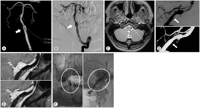
Fig. 2 A : Computed tomography angiography indicates a dissecting aneurysm at the intradural segment of the left vertebral artery. The length of the dilated segment was 7.5 mm (the arrow indicates the aneurysm). B : Digital subtraction angiography confirms the presence of a dissecting aneurysm at the same level with proximal stenosis (the arrow indicates the aneurysm). C : An axial image with a proton-density sequence at the level of the vertebral artery dissecting aneurysm obtained through high-resolution magnetic resonance imaging (HRMRI). D : Three-dimensional rotation angiography and digital subtraction angiography of the left vertebral artery show a dissecting aneurysm (the arrow indicates the real inlet of the dissecting segment). E : HRMRI shows the hidden pseudolumen. The length of the hidden pseudolumen was 7 mm, compared with that noted on digital subtraction angiography. Furthermore, the hidden pseudolumen and intramural hematoma were located on the opposite side of the dissecting aneurysm that was identified on angiography. F : Angiography after stent-assisted coiling in the working angle indicates a relatively well-preserved vertebral artery.
Cited by 1 articles
-
Vertebral Artery Dissecting Aneurysm Causing Central Tapia’s Syndrome: A Case Report
Yong Woo Shim, Jung Hyun Park, Sung-Tae Kim, Jin Wook Baek, Hyun Gon Lee, Jung Hae Ko, Sung Hwa Paeng, Se Young Pyo, Sung-Chul Jin, Hae Woong Jeong, Young Gyun Jeong
Neurointervention. 2021;16(2):185-189. doi: 10.5469/neuroint.2021.00080.
Reference
-
1. Andoh T, Shirakami S, Nakashima T, Nishimura Y, Sakai N, Yamada H, et al. Clinical analysis of a series of vertebral aneurysm cases. Neurosurgery. 1992; 31:987–993. discussion 993PMID: 1470333.
Article2. Anxionnat R, de Melo Neto JF, Bracard S, Lacour JC, Pinelli C, Civit T, et al. Treatment of hemorrhagic intracranial dissections. Neurosurgery. 2003; 53:289–300. discussion 300-301PMID: 12925243.
Article3. Bachmann R, Nassenstein I, Kooijman H, Dittrich R, Stehling C, Kugel H, et al. High-resolution magnetic resonance imaging (MRI) at 3.0 Tesla in the short-term follow-up of patients with proven cervical artery dissection. Invest Radiol. 2007; 42:460–466. PMID: 17507819.
Article4. Bodle JD, Feldmann E, Swartz RH, Rumboldt Z, Brown T, Turan TN. High-resolution magnetic resonance imaging : an emerging tool for evaluating intracranial arterial disease. Stroke. 2013; 44:287–292. PMID: 23204050.5. Chung JW, Kim BJ, Choi BS, Sohn CH, Bae H, Yoon BW, et al. High-resolution magnetic resonance imaging reveals hidden etiologies of symptomatic vertebral arterial lesions. J Stroke Cerebrovasc Dis. 2014; 23:293–302. PMID: 23541422.
Article6. Hunter MA, Santosh C, Teasdale E, Forbes KP. High-resolution double inversion recovery black-blood imaging of cervical artery dissection using 3T MR imaging. AJNR Am J Neuroradiol. 2012; 33:E133–E137. PMID: 21852374.
Article7. Jiang WJ, Yu W, Ma N, Du B, Lou X, Rasmussen PA. High resolution MRI guided endovascular intervention of basilar artery disease. J Neurointerv Surg. 2011; 3:375–378. PMID: 21990448.
Article8. Kai Y, Nishi T, Watanabe M, Morioka M, Hirano T, Yano S, et al. Strategy for treating unruptured vertebral artery dissecting aneurysms. Neurosurgery. 2011; 69:1085–1091. discussion 1091-1092PMID: 21629133.
Article9. Kwak HS, Hwang SB, Chung GH, Jeong SK. High-resolution magnetic resonance imaging of symptomatic middle cerebral artery dissection. J Stroke Cerebrovasc Dis. 2014; 23:550–553. PMID: 23635923.
Article10. Lee JM, Kim TS, Joo SP, Yoon W, Choi HY. Endovascular treatment of ruptured dissecting vertebral artery aneurysms--long-term follow-up results, benefits of early embolization, and predictors of outcome. Acta Neurochir (Wien). 2010; 152:1455–1465. PMID: 20467760.
Article11. Mizutani T. A fatal, chronically growing basilar artery : a new type of dissecting aneurysm. J Neurosurg. 1996; 84:962–971. PMID: 8847591.
Article12. Mizutani T. Natural course of intracranial arterial dissections. J Neurosurg. 2011; 114:1037–1044. PMID: 20950090.
Article13. Mizutani T, Kojima H, Asamoto S. Healing process for cerebral dissecting aneurysms presenting with subarachnoid hemorrhage. Neurosurgery. 2004; 54:342–347. discussion 347-348PMID: 14744280.
Article14. Naito I, Iwai T, Sasaki T. Management of intracranial vertebral artery dissections initially presenting without subarachnoid hemorrhage. Neurosurgery. 2002; 51:930–937. discussion 937-938PMID: 12234399.
Article15. Nakagawa K, Touho H, Morisako T, Osaka Y, Tatsuzawa K, Nakae H, et al. Long-term follow-up study of unruptured vertebral artery dissection : clinical outcomes and serial angiographic findings. J Neurosurg. 2000; 93:19–25. PMID: 10883900.
Article16. Peluso JP, van Rooij WJ, Sluzewski M, Beute GN, Majoie CB. Endovascular treatment of symptomatic intradural vertebral dissecting aneurysms. AJNR Am J Neuroradiol. 2008; 29:102–106. PMID: 17928377.
Article17. Roccatagliata L, Guédin P, Condette-Auliac S, Gaillard S, Colas F, Boulin A, et al. Partially thrombosed intracranial aneurysms : symptoms, evolution, and therapeutic management. Acta Neurochir (Wien). 2010; 152:2133–2142. PMID: 20725843.
Article18. Ryu CW, Kwak HS, Jahng GH, Lee HN. High-resolution MRI of intracranial atherosclerotic disease. Neurointervention. 2014; 9:9–20. PMID: 24644529.
Article
- Full Text Links
-
- Actions
-
Cited
- CITED
-
- Close
- Share
-
- Similar articles
-
- Two Cases of Intracranial Vertebral Artery Dissecting Aneurysm Improved by Antiplatelets Therapy
- Spontaneous Resolution of Dissecting Aneurysm of the Vertebral Artery
- Dissecting Aneurysm of the Intracranial Vertbral Artery: Case Report
- Nontraumatic Intracranial Dissecting Aneurysm of Vertebral Artery: Case Report
- Stent Assisted Coil Embolization of a Dissecting Aneurysm of the Vertebral Artery: A Case Involving a Patient with Hypoplasia of the Contralateral Vertebral Artery
