Changes in Tear Meniscus Height Following Lower Blepharoplasty as Measured by Optical Coherence Tomography
- Affiliations
-
- 1Department of Ophthalmology, CHA Bundang Medical Center, CHA University, Seongnam, Korea. eye@cha.ac.kr
- KMID: 2422063
- DOI: http://doi.org/10.3341/kjo.2017.0125
Abstract
- PURPOSE
The lower eyelid serves important roles in tear distribution and drainage. The purpose of this study was to measure the tear meniscus height (TMH) with anterior segment optical coherence tomography after lower blepharoplasty.
METHODS
A total of 52 eyes from 26 patients treated between July 2012 and June 2015 were included in the study. A transcutaneous or transconjunctival approach was performed, depending on whether (1) the supportive lower lid orbicularis oculi muscle was tightened, (2) the middle lamella was elongated, (3) minimal fat was removed or transpositioned, and (4) lateral canthal support was established. Marginal reflex distance 2 and marginal nose distance were analyzed with Image J software. TMH was measured with anterior segment optical coherence tomography. A paired t-test and Wilcoxon signed-rank test were used for statistical comparisons.
RESULTS
Marginal reflex distance 2 decreased and marginal nose distance increased with both surgical techniques. TMH decreased from 337.3 ± 117.9 to 289.3 ± 69.1 µm (p = 0.024) in patients who had transcutaneous lower blepharoplasty, but increased from 186.5 ± 35.5 to 274.8 ± 58.3 µm (p = 0.000) in patients who had transconjunctival lower blepharoplasty. Medial and lateral TMHs decreased significantly from 228.8 ± 80.7 to 152.7 ± 42.1 µm (p = 0.008) in patients with transcutaneous lower blepharoplasty. TMH was significantly restored after lower blepharoplasty with either approach.
CONCLUSIONS
Enhancing the lower eyelid position combined with orbicularis muscle tightening and lateral canthal support can normalize the TMH following lower eyelid blepharoplasty.
Keyword
Figure
-
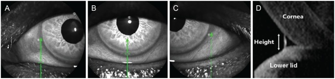
Fig. 1 Lower eyelid tear meniscus height was measured by anterior segment optical coherence tomography with a Spectralis optical coherence tomography. (A) Medial point, (B) central point, (C) lateral point, and (D) cross-sectional view. Green arrows indicate the vertical line centered at three points (medial, central, and lateral).
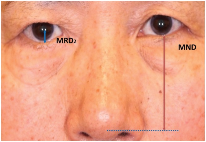
Fig. 2 Measurement of clinical parameters of lower eyelid position. Marginal reflex distance 2 (MRD2) and marginal nose distance (MND) were analyzed with ImageJ software.

Fig. 3 Surgical procedures used in lower blepharoplasty. (A–C) The transcutaneous approach. (A) An incision was made 1 mm below the lower eyelid margin, (B) followed by incision of the orbital septum, and (C) removal of orbital fat. (D–F) The transconjunctival approach. (D) An incision was made at the conjunctiva 3 to 4 mm below the tarsal plate, (E) followed by an incision at the orbital septum, and (F) removal of orbital fat. (G–I) Lateral canthal tightening procedures, (G) orbicularis muscle tightening and lateral canthopexy, (H) lateral canthal suspension, and (I) lateral tarsal strip.

Fig. 4 The changes of lower eyelid tear meniscus height after lower blepharoplasty. (A) Transcutaneous approach (n = 34, Wilcoxon signed-rank test; *p < 0.05), (B) transconjunctival approach (n = 18, Wilcoxon signed-rank test; *p < 0.05), (C) total patients (Wilcoxon signed-rank test, *p < 0.05).
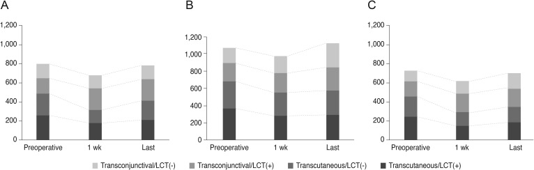
Fig. 5 Lower eyelid tear meniscus height at three points (A, medial; B, central; C, lateral) after lower blepharoplasty with all lateral canthal tightening (LCT) procedures.
Reference
-
1. Buchanan DR, Wulc AE. Contemporary thoughts on lower eyelid/midface aging. Clin Plast Surg. 2015; 42:1–15. PMID: 25440737.
Article2. Tse DT, Erickson BP, Tse BC. The BLICK mnemonic for clinical-anatomical assessment of patients with epiphora. Ophthalmic Plast Reconstr Surg. 2014; 30:450–458. PMID: 25216202.
Article3. Prischmann J, Sufyan A, Ting JY, et al. Dry eye symptoms and chemosis following blepharoplasty: a 10-year retrospective review of 892 cases in a single-surgeon series. JAMA Facial Plast Surg. 2013; 15:39–46. PMID: 23329270.4. Honrado CP, Pastorek NJ. Long-term results of lower-lid suspension blepharoplasty: a 30-year experience. Arch Facial Plast Surg. 2004; 6:150–154. PMID: 15148120.5. Hamawy AH, Farkas JP, Fagien S, Rohrich RJ. Preventing and managing dry eyes after periorbital surgery: a retrospective review. Plast Reconstr Surg. 2009; 123:353–359. PMID: 19116572.
Article6. Shao C, Fu Y, Lu L, et al. Dynamic changes of tear fluid after cosmetic transcutaneous lower blepharoplasty measured by optical coherence tomography. Am J Ophthalmol. 2014; 158:55–63. PMID: 24709809.
Article7. Huang D, Swanson EA, Lin CP, et al. Optical coherence tomography. Science. 1991; 254:1178–1181. PMID: 1957169.
Article8. Wang J, Aquavella J, Palakuru J, et al. Relationships between central tear film thickness and tear menisci of the upper and lower eyelids. Invest Ophthalmol Vis Sci. 2006; 47:4349–4355. PMID: 17003425.
Article9. Wang Y, Zhuang H, Xu J, et al. Dynamic changes in the lower tear meniscus after instillation of artificial tears. Cornea. 2010; 29:404–408. PMID: 20164745.
Article10. Mainstone JC, Bruce AS, Golding TR. Tear meniscus measurement in the diagnosis of dry eye. Curr Eye Res. 1996; 15:653–661. PMID: 8670769.
Article11. Park DI, Lew H, Lee SY. Tear meniscus measurement in nasolacrimal duct obstruction patients with Fourier-domain optical coherence tomography: novel three-point capture method. Acta Ophthalmol. 2012; 90:783–787. PMID: 21726426.
Article12. Robinson FO, Collin RO. Ectropion. In : Yanoff M, Duker JS, editors. Ophthalmology. 2nd ed. Edinburgh: Mosby;2006. p. 676–683.13. Taban M, Taban M, Perry JD. Lower eyelid position after transconjunctival lower blepharoplasty with versus without a skin pinch. Ophthalmic Plast Reconstr Surg. 2008; 24:7–9. PMID: 18209632.
Article14. Sultan B, Genther DJ, Perkins SW. Measurement of change in lower eyelid position in patients undergoing transcutaneous skin-muscle flap lower eyelid blepharoplasty. JAMA Facial Plast Surg. 2016; 18:429–435. PMID: 27390027.
Article15. Segal KL, Patel P, Levine B, et al. The effect of transconjunctival blepharoplasty on margin reflex distance 2. Aesthetic Plast Surg. 2016; 40:13–18. PMID: 26537512.
Article16. Sung YJ, Park JS, Lew H. Changes in lower eyelid positions after individualized lower blepharoplasty. J Korean Ophthalmol Soc. 2015; 56:1831–1839.
Article17. Zhou S, Li Y, Lu AT, et al. Reproducibility of tear meniscus measurement by Fourier-domain optical coherence tomography: a pilot study. Ophthalmic Surg Lasers Imaging. 2009; 40:442–447. PMID: 19772266.
Article18. Palakuru JR, Wang J, Aquavella JV. Effect of blinking on tear dynamics. Invest Ophthalmol Vis Sci. 2007; 48:3032–3037. PMID: 17591869.
Article19. Wang J, Aquavella J, Palakuru J, Chung S. Repeated measurements of dynamic tear distribution on the ocular surface after instillation of artificial tears. Invest Ophthalmol Vis Sci. 2006; 47:3325–3329. PMID: 16877398.
Article20. Johnson ME, Murphy PJ. The agreement and repeatability of tear meniscus height measurement methods. Optom Vis Sci. 2005; 82:1030–1037. PMID: 16357644.
Article21. Shen M, Li J, Wang J, et al. Upper and lower tear menisci in the diagnosis of dry eye. Invest Ophthalmol Vis Sci. 2009; 50:2722–2726. PMID: 19218609.
Article
- Full Text Links
-
- Actions
-
Cited
- CITED
-
- Close
- Share
-
- Similar articles
-
- Tear Meniscus Evaluation Using Optical Coherence Tomography in Dry Eye Patients
- Changes in Area of Conjunctiva and Tear Meniscus Measured Using Anterior Segment Optical Coherence Tomography after Conjunctivochalasis Surgery
- Analysis of Tear Meniscus Change after Strabismus Surgery Using Optical Coherence Tomography
- Repeatability and Reproducibility of Tear Meniscus Evaluations Using Two Different Spectral Domain-optical Coherence Tomography
- Tear Meniscus Evaluation Using Optical Coherence Tomography in Meibomein Gland Dysfunction Patients
