Hydrocephalus as a Complication of Durotomy during Cervical Laminoplasty: A Case Report
- Affiliations
-
- 1Department of Orthopaedic Surgery, Dankook University College of Medicine, Cheonan, Korea. firekimdo@gmail.com
- KMID: 2421613
- DOI: http://doi.org/10.4184/jkss.2018.25.2.69
Abstract
- STUDY DESIGN: Case report.
OBJECTIVES
We report a case of hydrocephalus as a complication of durotomy during cervical laminoplasty. SUMMARY OF LITERATURE REVIEW: Hydrocephalus is a very rare complication of cervical laminoplasty.
MATERIALS AND METHODS
A 72-year-old man had an incidental durotomy during cervical laminoplasty. The dural leak was repaired by secondary surgery. However, the patient continued to complain of headaches and developed confusion and drowsiness. A computed tomographic scan of the brain showed hydrocephalus. After insertion of a lumbar drain, the patient experienced a temporary improvement in the neurologic symptoms. After 6 months, the neurologic symptoms recurred and a ventriculoperitoneal (VP) shunt was placed.
RESULTS
After placement of the VP shunt, the neurologic symptoms improved significantly.
CONCLUSIONS
If a patient shows deterioration of neurologic symptoms after an incidental durotomy, surgeons should consider the possibility of hydrocephalus.
Keyword
MeSH Terms
Figure
-
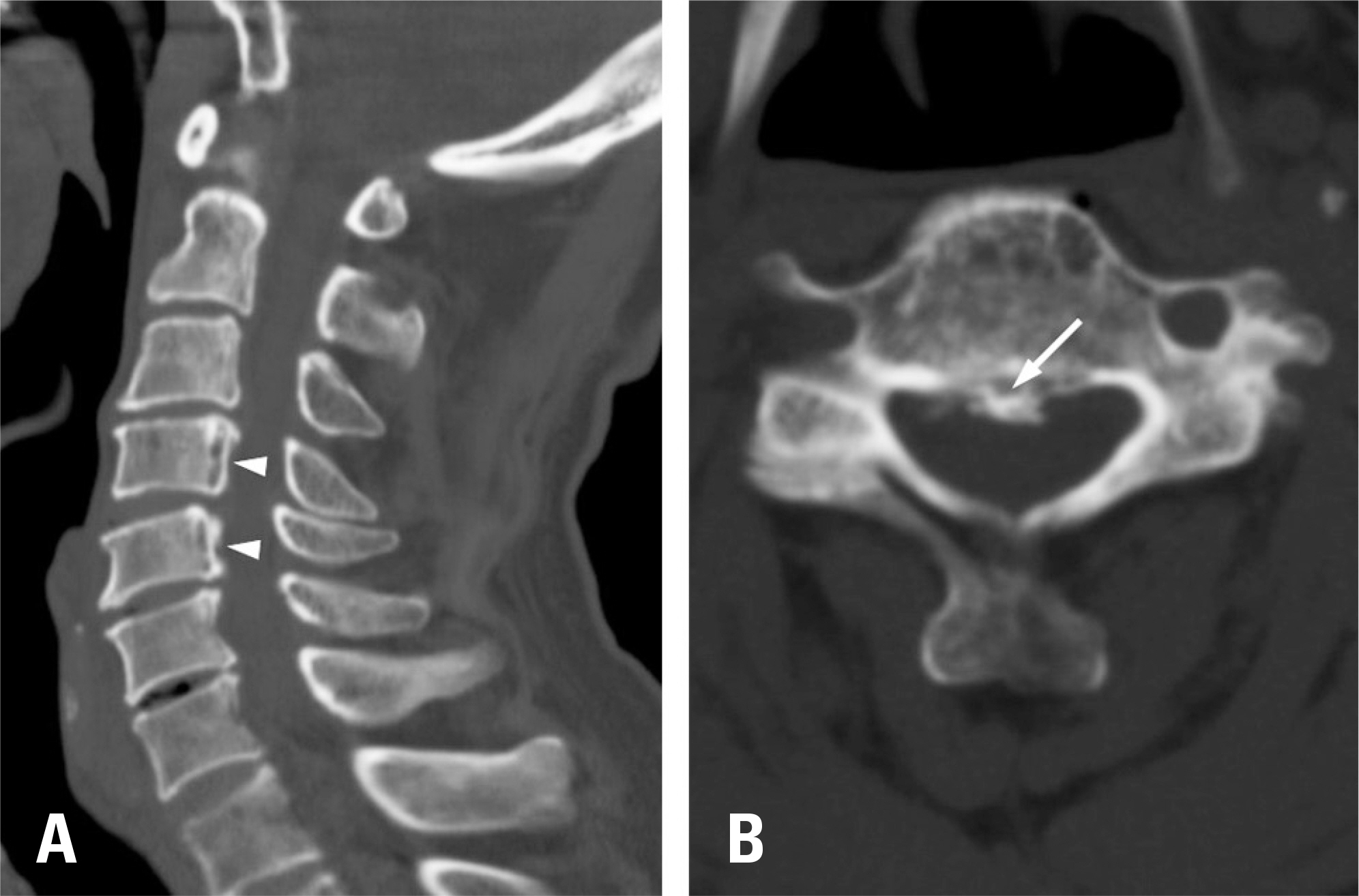
Fig. 1. Preoperative computed tomographic (CT) scan of the cervical spine. (A) A sagittal CT scan shows segmental ossification of the posterior longitudinal ligament (OPLL, arrowheads) spanning from C4 to C5. (B) An axial CT scan at C4 shows encroachment of the spinal canal by the OPLL mass (arrow).
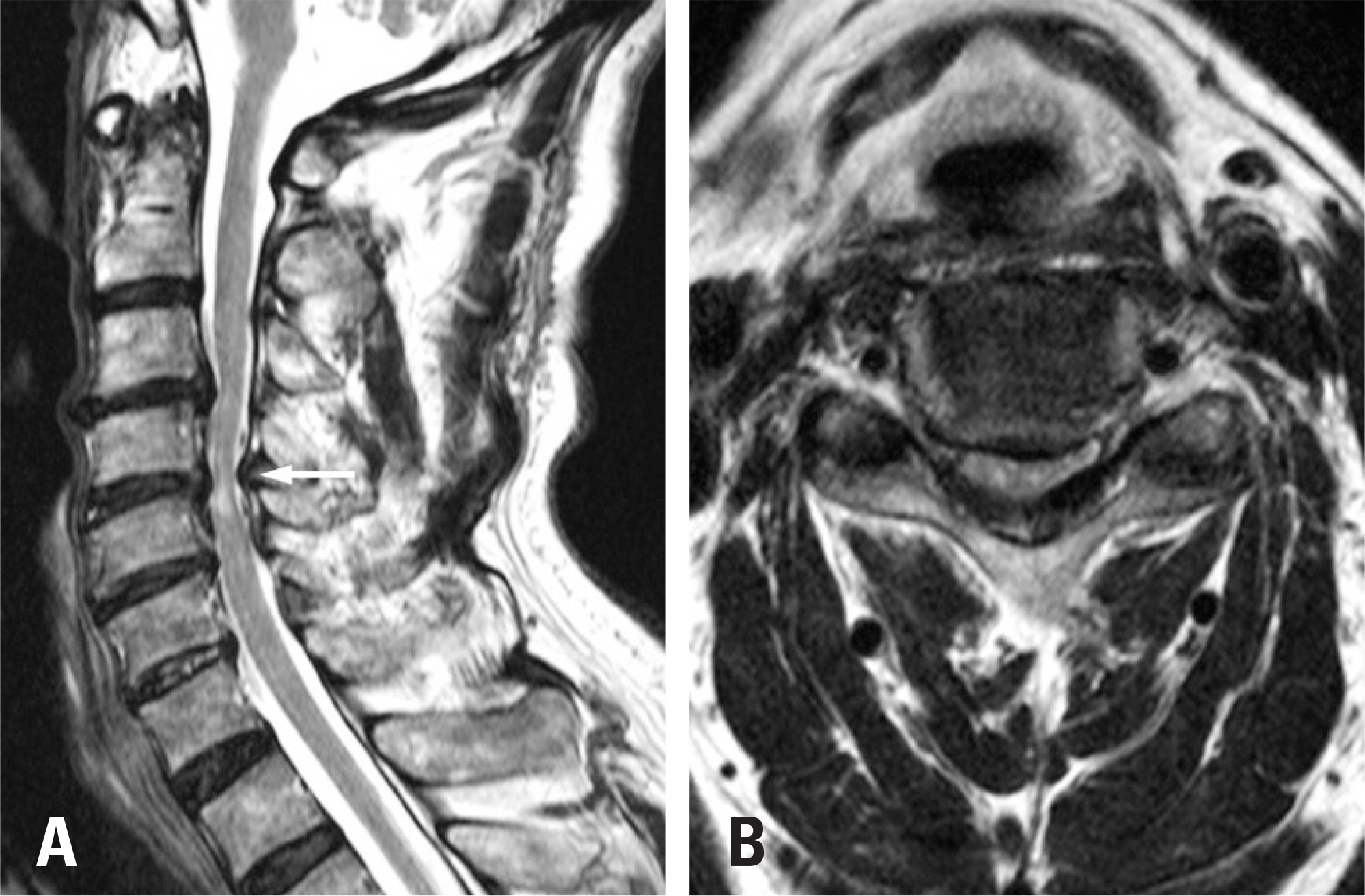
Fig. 2. Preoperative T2-weighted magnetic resonance imaging (MRI) scan of the cervical spine. (A) A sagittal MRI scan shows increased cord signal intensity (arrow) at C4-C5. (B) An axial MRI scan shows central canal stenosis at C4-C5.
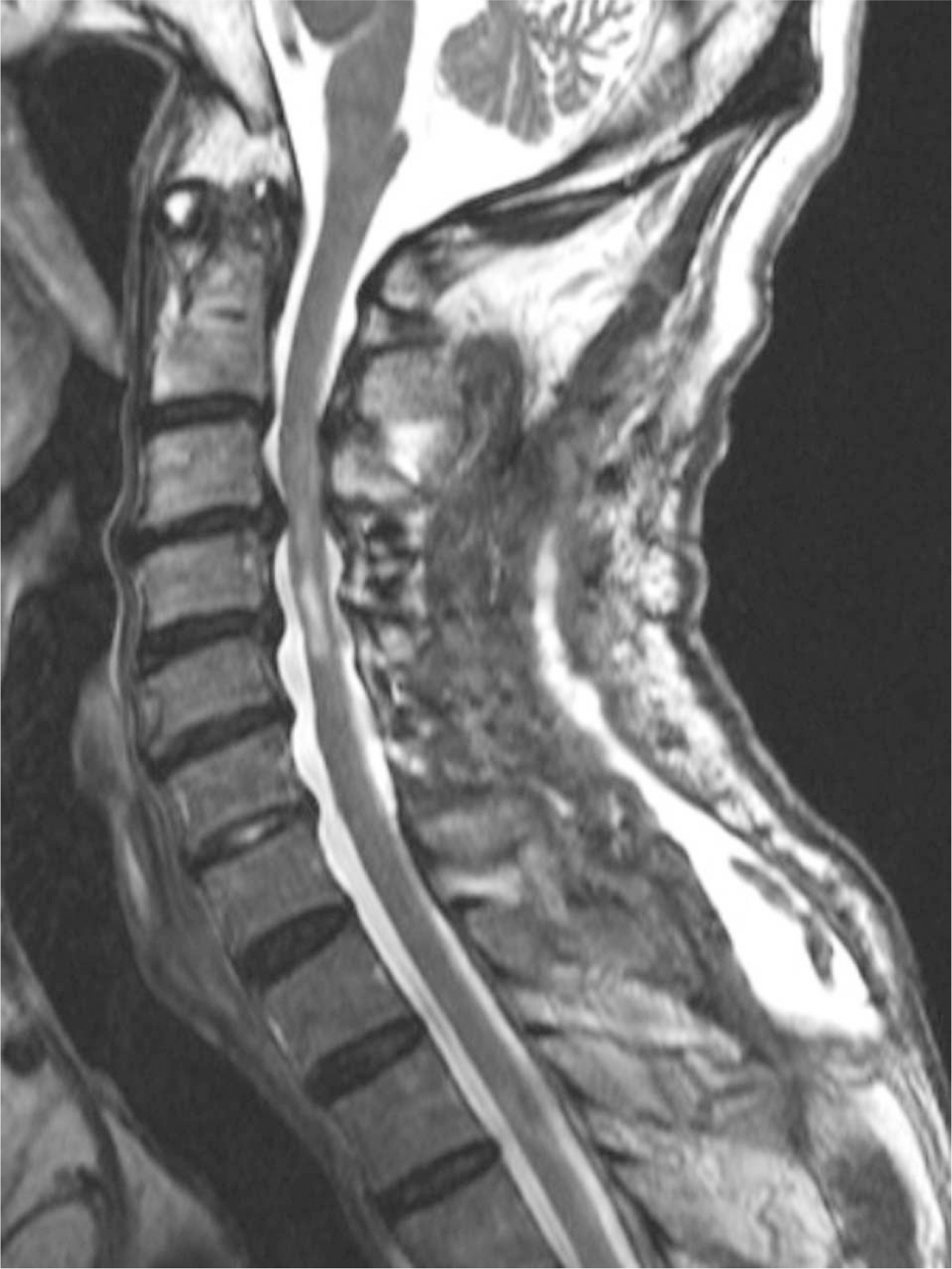
Fig. 3. T2-weighted sagittal magnetic resonance imaging shows adequate decompression and subcutaneous fluid collection.

Fig. 4. Computed tomography shows lateral ventricular dilation.

Fig. 5. Computed tomography 30 days after ventriculoperitoneal shunt placement shows normal ventricular morphology.
Reference
-
1. Wada E, Yonenobu K. Treatment of cervical myelopathy: Laminoplasty. Benzel EC, editor. eds.The cervical spine 5ed. New York: Lippincott Williams & Wilkins;2012. 980-94.2. Mirone G, Cinalli G, Spennato P, et al. Hydrocephalus and spinal cord tumors: a review. Childs Nerv Syst. 2011 Oct; 27(10):1741–9. DOI: 10.1007/s00381-011-1543-5.
Article3. Joseph G, Johnston RA, Fraser MH, et al. Delayed hydrocephalus as an unusual complication of a stab injury to the spine. Spinal Cord. 2005 Jan; 43(1):56–8. DOI: 10.1038/sj.sc.3101655.
Article4. Matsuda R, Goda K, Nakase H, et al. Case of hydrocephalus after cervical laminoplasty for cervical ossification of the posterior longitudinal ligament. Brain Nerve. 2009 Jan; 61(1):89–92.5. Matsushima K, Hashimoto R, Gondo M, et al. Perifascial Areolar Tissue Graft for Spinal Dural Repair with Cerebrospinal Fluid Leakage: Case Report of Novel Graft Material, Radiological Assessment Technique, and Rare Postoperative Hydrocephalus. World Neurosurgery. 2016 Nov; 95:619. DOI: 10.1016/j.wneu.2016.08.025.
Article6. Maezawa Y, Baba H, Annen S, et al. Development of hydrocephalus after cervical laminoplasty for ossification of the posterior longitudinal ligament: case report. Spinal Cord. 1996 Nov; 34(11):699–702. DOI: DOI:10.1038/sc.1996.127.
Article7. Cavanilles-Walker JM, Tomasi SO, et al. Remote cerebellar haemorrhage after lumbar spine surgery: case report. Arch Orthop Trauma Surg. 2013 Dec; 133(12):1645–8. DOI: 10.1007/s00402-013-1867-6.
Article8. Endriga DT, Dimar JR 2nd, Carreon LY. Communicating hydrocephalus, a long-term complication of dural tear during lumbar spine surgery. Eur Spine J. 2016 May; 25(1 Suppl):157–61. DOI: 10.1007/s00586-015-4308-0.
Article9. Koerts G, Rooijakkers H, Abu-Serieh B, et al. Postoperative spinal adhesive arachnoiditis presenting with hydrocephalus and cauda equine syndrome. Clin Neurol Neurosurg. 2008 Feb; 110(2):171–5. DOI: 10.1016/j.clineuro.2007.09.004.10. Zeidman SM. Cervical cerebrospinal fluid leakage, durotomy, and pseudomeningocele. Benzel EC, editor. eds.The cervical spine 5ed. New York: Lippincott Williams & Wilkins;2012. 1294-300.
- Full Text Links
-
- Actions
-
Cited
- CITED
-
- Close
- Share
-
- Similar articles
-
- New Cervical Laminoplasty Polyethererketone Cage: Two Case Reports
- Acute Hydrocephalus as a Complication of Cervical Spine Fracture and Dislocation: A Case Report
- Laminoplasty Versus Laminectomy and Fusion for Multilevel Cervical Spondylosis
- Expansive Laminoplasty for Cervical Compression Myelopathy
- The Usefulness of Laminoplasty in Cervical Spinal Cord Tumor Surgery
