Anat Cell Biol.
2018 Jun;51(2):136-138. 10.5115/acb.2018.51.2.136.
Solitary osteochondroma in the body of the pubic bone: a cadaveric case report
- Affiliations
-
- 1Department of Anatomy, Melaka Manipal Medical College (Manipal Campus), Manipal Academy of Higher Education, Manipal, India.
- 2Department of Human and Clinical Anatomy, College of Medicine and Health Sciences, Sultan Qaboos University, Muscat, Oman. srinivasa@squ.edu.om
- 3Department of Pathology, Kasturba Medical College, Manipal Academy of Higher Education, Manipal, India.
- 4Faculty in Anatomy, Jeffrey Cheah School of Medicine & Health Sciences, Monash University, Sunway Campus, Kuala Lumpur, Malaysia.
- KMID: 2421178
- DOI: http://doi.org/10.5115/acb.2018.51.2.136
Abstract
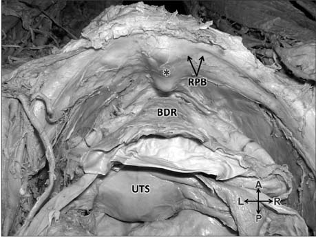
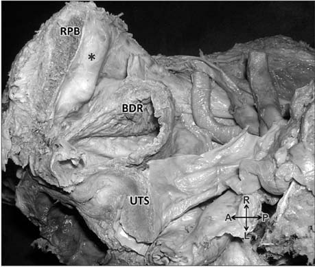
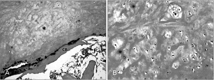
- Osteochondromas develop as cartilaginous nodules in the periosteum of bones. They are the commonest benign tumors of the skeleton, generally observed in the long bones. Rarely, they are also found in the axial skeleton, flat bones of skull and facial bones. During a regular dissection, we came across a solitary osteochondroma in posterior surface of the body of the right pubic bone. Histopathology of the bony projection confirmed the typical features of the osteochondroma. The symptomatic osteochondromas are usually evaluated during radiographic examination. Though, the observed osteochondroma is relatively smaller its unusual location is remarkable and knowledge of occurrence of such nodules is clinically important during the diagnosis and planning of treatment.
Keyword
Figure
Reference
-
1. Murphey MD, Choi JJ, Kransdorf MJ, Flemming DJ, Gannon FH. Imaging of osteochondroma: variants and complications with radiologic-pathologic correlation. Radiographics. 2000; 20:1407–1434.
Article2. Price CH. Primary bone-forming tumours and their relationship to skeletal growth. J Bone Joint Surg Br. 1958; 40-B:574–593.
Article3. Strange FG. Excision of the superior ramus of the pubis for large osteochondroma. Br J Surg. 1954; 41:377–379.4. Kumar S, Shah AK, Patel AM, Shah UA. CT and MR images of flat bone osteochondromata from head to foot: a pictorial essay. Indian J Radiol Imaging. 2006; 16:589–596.5. Hoshimoto K, Mitsuya K, Ohkura T. Osteochondroma of the pubic symphysis associated with sexual disturbance. Gynecol Obstet Invest. 2000; 50:70–72.
Article6. Buzon MR. Two cases of pelvic osteochondroma in New Kingdom Nubia. Int J Osteoarchaeol. 2005; 15:377–382.
Article7. Mnif H, Zrig M, Koubaa M, Zammel N, Abid A. An unusual complication of pubic exostosis. Orthop Traumatol Surg Res. 2009; 95:151–153.
Article8. Herode P, Shroff A, Patel P, Aggarwal P, Mandlewala V. A rare case of pubic ramus osteochondroma. J Orthop Case Rep. 2015; 5:51–53.9. Vallance R, Hamblen DL, Kelly IG. Vascular complications of osteochondroma. Clin Radiol. 1985; 36:639–642.
Article10. Karasick D, Schweitzer ME, Eschelman DJ. Symptomatic osteochondromas: imaging features. AJR Am J Roentgenol. 1997; 168:1507–1512.
Article
- Full Text Links
- Actions
-
Cited
- CITED
-
- Close
- Share
- Similar articles
-
- Solitary Senescent Osteochondroma of the Sacrum Producing Sciatica: A Case Report
- Osteochondroma of the Sacrum: A Case Report
- Arthroscopic Excision of Intra-articular Osteochondroma of the Elbow: A Case Report
- Case Report of Osteochondroma on the Mandible Body Area
- Solitary Osteochondroma of the Clavicle associated with Congenital Muscular Torticollis: A Case Report


