The relationship between masseter muscle thickness measured by ultrasonography and facial profile in young Korean adults
- Affiliations
-
- 1Department of Advanced General Dentistry, College of Dentistry, Yonsei University, Seoul, Korea. kdkim@yuhs.ac
- 2Seoul Dental Hospital for the Disabled, Seoul, Korea.
- 3School of Stomatology, Shandong University, Jinan Shi, Shandong Province, China.
- KMID: 2420549
- DOI: http://doi.org/10.5624/isd.2018.48.3.213
Abstract
- PURPOSE
The purpose of this study was to evaluate the relationship between masseter muscle thickness, facial morphology, and mandibular morphology in Korean adults using ultrasonography.
MATERIALS AND METHODS
Ultrasonography was used to measure the masseter muscle thickness bilaterally of 40 adults (20 males, 20 females) and was performed in the relaxed and contracted states. Facial photos and panoramic radiography were used for morphological analyses and evaluated for correlations with masseter muscle thickness. We also evaluated the correlations of age, body weight, stature, and body constitution with masseter muscle thickness.
RESULTS
In the relaxing, the masseter was 9.8±1.3 mm in females and 11.3±1.2 mm in males. In the contracted state, it was 12.4±1.4 mm in females and 14.7±1.4 mm in males. Facial photography showed that bizygomatic facial width over facial height was correlated with masseter muscle thickness in both sexes in the relaxed state, and was statistically significantly correlated with masseter muscle thickness in males in the contracted state. In panoramic radiography, correlations were found between anterior angle length and posterior angle length and masseter muscle thickness in females, and between body length and posterior angle length, between anterior angle length and body length, between ramal length and body length, and between body length and condyle length in males.
CONCLUSION
Masseter muscle thickness was associated with facial and mandibular morphology in both sexes, and with age in males. Ultrasonography can be used effectively to measure masseter muscle thickness.
MeSH Terms
Figure
-
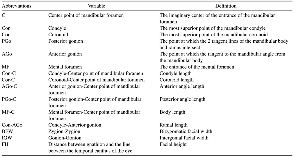
Fig. 1 Scanning level of the masseter muscle in axial imaging (yellow area).
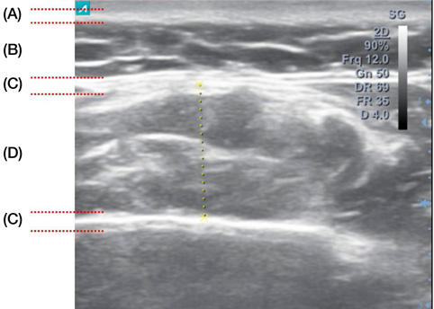
Fig. 2 Anatomical structure of masseter muscle in an ultrasound image. (A) skin layer; (B) fat layer; (C) masseteric fascia; (D) masseter muscle.
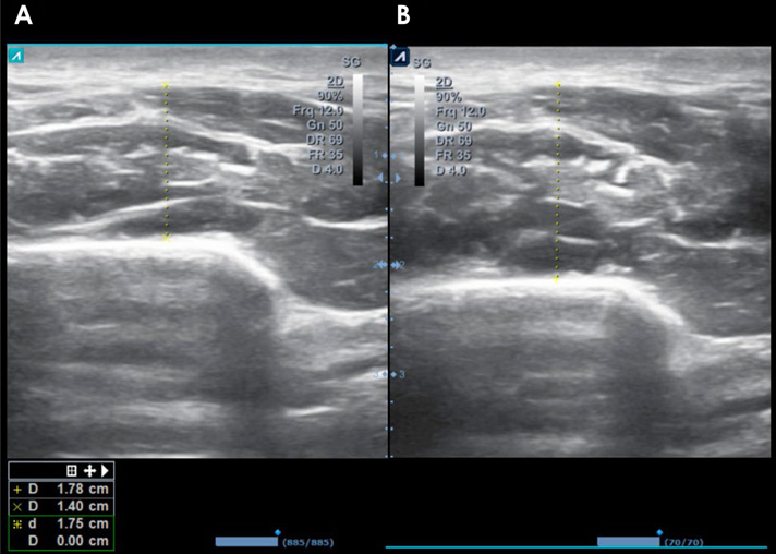
Fig. 3 Cross-sectional view of an ultrasound image. A. Relaxed state. B. Contracted state. The yellow dotted line is the masseter muscle thickness.

Fig. 4 Schematic diagram of reference points and measurements of length on a facial photograph. Bizygomatic facial width (BFW); intergonial facial width (IGW); facial height (FH).
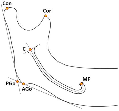
Fig. 5 Schematic diagram of reference points and measurements of length on a panoramic radiograph. Center point of mandibular foramen (C); condyle (Con); coronoid (Cor); posterior gonion (PGo); anterior gonion (AGo); mental foramen (MF).
Cited by 1 articles
-
Correlation between mandibular morphology and masticatory muscle thickness in normal occlusion and mandibular prognathism
Tae-Ho Kim, Chul-Hwan Kim
J Korean Assoc Oral Maxillofac Surg. 2020;46(5):313-320. doi: 10.5125/jkaoms.2020.46.5.313.
Reference
-
1. Ingervall B, Bitsanis E. A pilot study of the effect of masticatory muscle training on facial growth in long-face children. Eur J Orthod. 1987; 9:15–23.
Article2. Proffit WR, Fields HW, Nixon WL. Occlusal forces in normal-and long-face adults. J Dent Res. 1983; 62:566–570.3. Moss ML, Rankow RM. The role of the functional matrix in mandibular growth. Angle Orthod. 1968; 38:95–103.4. Moss ML, Simon MR. Growth of the human mandibular angular process: a functional cranial analysis. Am J Phys Anthropol. 1968; 28:127–138.
Article5. Moss ML, Salentijn L. The primary role of functional matrices in facial growth. Am J Orthod. 1969; 55:566–577.
Article6. Moss ML. Twenty years of functional cranial analysis. Am J Orthod. 1972; 61:479–485.
Article7. Uchida Y, Motoyoshi M, Shigeeda T, Shinohara A, Igarashi Y, Sakaguchi M, et al. Relationship between masseter muscle size and maxillary morphology. Eur J Orthod. 2011; 33:654–659.
Article8. Kubota M, Nakano H, Sanjo I, Satoh K, Sanjo T, Kamegai T, et al. Maxillofacial morphology and masseter muscle thickness in adults. Eur J Orthod. 1998; 20:535–542.
Article9. Park W, Kim BC, Yu HS, Yi CK, Lee SH. Architectural characteristics of the normal and deformity mandible revealed by three-dimensional functional unit analysis. Clin Oral Investig. 2010; 14:691–698.
Article10. Raadsheer MC, Van Eijden TM, Van Spronsen PH, Van Ginkel FC, Kiliaridis S, Prahl-Andersen B. A comparison of human masseter muscle thickness measured by ultrasonography and magnetic resonance imaging. Arch Oral Biol. 1994; 39:1079–1084.
Article11. Ariji Y, Kimura Y, Gotoh M, Sakuma S, Zhao YP, Ariji E. Blood flow in and around the masseter muscle: normal and pathologic features demonstrated by color Doppler sonography. Oral Surg Oral Med Oral Pathol Oral Radiol Endod. 2001; 91:472–482.
Article12. Ariji Y, Sakuma S, Izumi M, Sasaki J, Kurita K, Ogi N, et al. Ultrasonographic features of the masseter muscle in female patients with temporomandibular disorder associated with myofascial pain. Oral Surg Oral Med Oral Pathol Oral Radiol Endod. 2004; 98:337–341.
Article13. Takagi Y, Kimura Y, Nakamura H, Sasaki M, Eguchi K, Nakamura T. Salivary gland ultrasonography: can it be an alternative to sialography as an imaging modality for Sjogren's syndrome? Ann Rheum Dis. 2010; 69:1321–1324.
Article14. Yonetsu K, Ikemura K. Ultrasonographic study of the relation of metastatic nodes to the carotid artery. Head Neck Surg. 1987; 9:279–283.
Article15. Kiliaridis S, Kalebo P. Masseter muscle thickness measured by ultrasonography and its relation to facial morphology. J Dent Res. 1991; 70:1262–1265.
Article16. Kiliaridis S, Mahboubi PH, Raadsheer MC, Katsaros C. Ultrasonographic thickness of the masseter muscle in growing individuals with unilateral crossbite. Angle Orthod. 2007; 77:607–611.
Article17. Satiroğlu F, Arun T, Işik F. Comparative data on facial morphology and muscle thickness using ultrasonography. Eur J Orthod. 2005; 27:562–567.18. Tircoveluri S, Singh JR, Rayapudi N, Karra A, Begum M, Challa P. Correlation of masseter muscle thickness and intermolar width - an ultrasonography study. J Int Oral Health. 2013; 5:28–34.19. Turpin DL. Growth and remodeling of the mandible in the Macaca mulatta monkey. Am J Orthod. 1968; 54:251–271.
Article20. Cicchetti DV, Nelson LD. Re-examining threats to the reliability and validity of putative brain-behavior relationships: new guidelines for assessing the effect of patients lost to follow-up. J Clin Exp Neuropsychol. 1994; 16:339–343.
Article21. Bakke M, Tuxen A, Vilmann P, Jensen BR, Vilmann A, Toft M. Ultrasound image of human masseter muscle related to bite force, electromyography, facial morphology, and occlusal factors. Scand J Dent Res. 1992; 100:164–171.
Article22. Cotti E, Campisi G, Garau V, Puddu G. A new technique for the study of periapical bone lesions: ultrasound real time imaging. Int Endod J. 2002; 35:148–152.
Article23. Caivano D, Bufalari A, Giorgi ME, Conti MB, Marchesi MC, Angeli G, et al. Imaging diagnosis–transesophageal ultrasound-guided removal of a migrating grass awn foreign body in a dog. Vet Radiol Ultrasound. 2014; 55:561–564.
Article24. Bruneton JN, Balu-Maestro C, Marcy PY, Melia P, Mourou MY. Very high frequency (13 MHz) ultrasonographic examination of the normal neck: detection of normal lymph nodes and thyroid nodules. J Ultrasound Med. 1994; 13:87–90.
Article25. Yasumoto M, Nakagawa T, Shibuya H, Suzuki S, Satoh T. Ultrasonography of the sublingual space. J Ultrasound Med. 1993; 12:723–729.
Article26. Ng TK, Vasilareas D, Mitterdorfer AJ, Maher PO, Lalak A. Prostate cancer detection with digital rectal examination, prostate-specific antigen, transrectal ultrasonography and biopsy in clinical urological practice. BJU Int. 2005; 95:545–548.
Article27. Savitha B, Vandana KL. Comparative assesment of gingival thickness using transgingival probing and ultrasonographic method. Indian J Dent Res. 2005; 16:135–139.
Article28. Aggarwal V, Logani A, Shah N. RETRACTED: The evaluation of computed tomography scans and ultrasounds in the differential diagnosis of periapical lesions. J Endod. 2008; 34:1312–1315.
Article
- Full Text Links
-
- Actions
-
Cited
- CITED
-
- Close
- Share
-
- Similar articles
-
- A Reproducible and Reliable Method for Measuring Masseter Muscle Thickness in Maximal Bite Force Using Ultrasonography
- An Ultrasonographic Evaluation of Masseter Muscle Thickness in Patients Having Parafunctional Habit
- Ultrasonographic study on the masseter muscle thickness of adult Korean
- Ultrasonographic evaluation of the masseter muscle in patients with temporomandibular joint degeneration
- Evaluation of normal masseter muscles on ultrasonography
