Influence of voxel size on cone-beam computed tomography-based detection of vertical root fractures in the presence of intracanal metallic posts
- Affiliations
-
- 1Department of Stomatologic Sciences, School of Dentistry, Federal University of Goiás, Goiânia, Brazil.
- 2Department of Oral Rehabilitation, Dental School, Federal University of Goiás, Goiânia, Brazil.
- 3Department of Oral Pathology, School of Dentistry, University of Anápolis, Anápolis, Brazil. brunno.santosfreitas@gmail.com
- KMID: 2420545
- DOI: http://doi.org/10.5624/isd.2018.48.3.177
Abstract
- PURPOSE
This study was performed to evaluate the influence of voxel size and the accuracy of 2 cone-beam computed tomography (CBCT) systems in the detection of vertical root fracture (VRF) in the presence of intracanal metallic posts.
MATERIALS AND METHODS
Thirty uniradicular extracted human teeth were selected and randomly divided into 2 groups (VRF group, n=15; and control group, n=15). The VRFs were induced by an Instron machine, and metallic posts were placed in both groups. The scans were acquired by CBCT with 4 different voxel sizes: 0.1 mm and 0.16 mm (for the Eagle 3D V-Beam system) and 0.125 mm and 0.2 mm (for the i-CAT system) (protocols 1, 2, 3, and 4, respectively). Interobserver and intraobserver agreement was assessed using the Cohen kappa test. Sensitivity and specificity were evaluated and receiver operating characteristic analysis was performed.
RESULTS
The intraobserver coefficients indicated good (0.71) to very good (0.83) agreement, and the interobserver coefficients indicated moderate (0.57) to very good (0.80) agreement. In respect to the relationship between sensitivity and specificity, a statistically significant difference was found between protocols 1 (positive predictive value: 0.710, negative predictive value: 0.724) and 3 (positive predictive value: 0.727, negative predictive value: 0.632) (P < .05). The least interference due to artifact formation was observed using protocol 2.
CONCLUSION
Protocols with a smaller voxel size and field of view seemed to favor the detection of VRF in teeth with intracanal metallic posts.
MeSH Terms
Figure
-
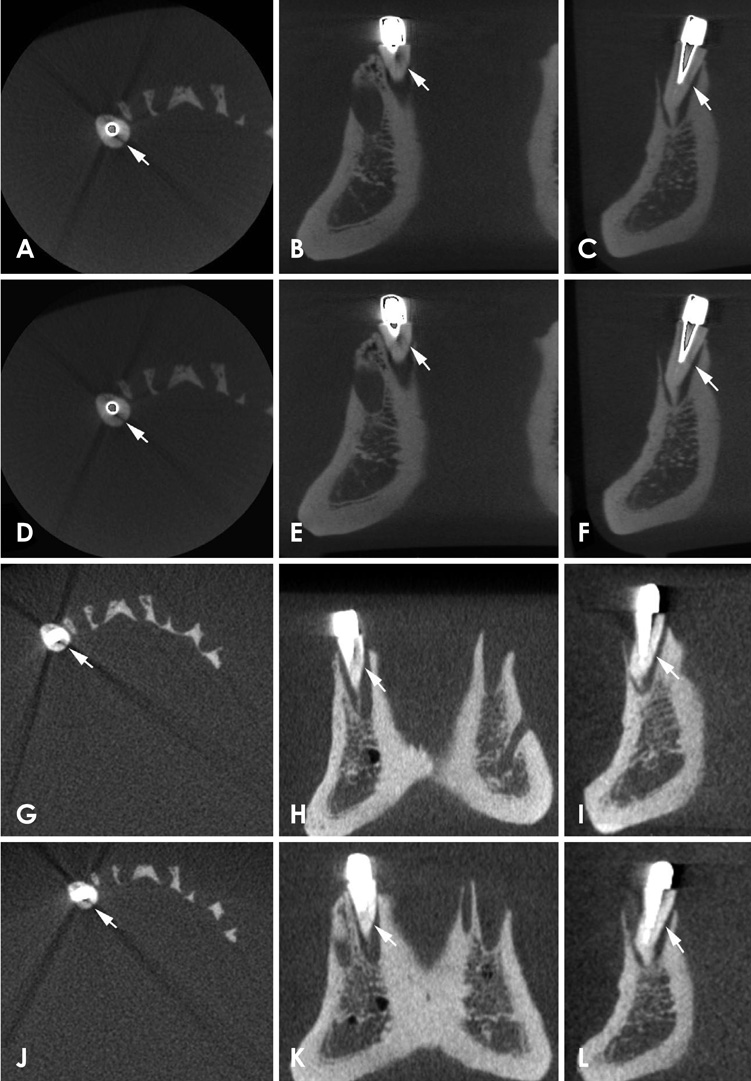
Fig. 1 Images of vertical root fracture with a metallic post in 2 cone-beam computed tomography systems with different voxel sizes. Protocol 1 with 0.1-mm voxels in (A) the axial plane (A), the coronal plane (B), and the cross-sectional plane (C). Protocol 2 with 0.16-mm voxels in the axial plane (D), the coronal plane (E), and the cross-sectional plane (F). Protocol 3 with 0.125-mm voxels in the axial plane (G), the coronal plane (H), and the cross-sectional plane (I). Protocol 4 with 0.2-mm voxels in the axial plane (J), the coronal plane (K), and the cross-sectional plane (L).

Fig. 2 Receiver operating characteristic curves of the scanning protocols and cone-beam computed tomography systems.
Cited by 1 articles
-
Influence of CBCT parameters on image quality and the diagnosis of vertical root fractures in teeth with metallic posts: an
ex vivo study
Larissa Pereira Lagos de Melo, Polyane Mazucatto Queiroz, Larissa Moreira-Souza, Mariana Rocha Nadaes, Gustavo Machado Santaella, Matheus Lima Oliveira, Deborah Queiroz Freitas
Restor Dent Endod. 2023;48(2):e16. doi: 10.5395/rde.2023.48.e16.
Reference
-
1. Chai H, Tamse A. The effect of isthmus on vertical root fracture in endodontically treated teeth. J Endod. 2015; 41:1515–1519.
Article2. Ferreira LM, Visconti MA, Nascimento HA, Dallemolle RR, Ambrosano GM, Freitas DQ. Influence of CBCT enhancement filters on diagnosis of vertical root fractures: a simulation study in endodontically treated teeth with and without intracanal posts. Dentomaxillofac Radiol. 2015; 44:20140352.
Article3. Fuss Z, Lustig J, Katz A, Tamse A. An evaluation of endodontically treated vertical root fractured teeth: impact of operative procedures. J Endod. 2001; 27:46–48.
Article4. Chang E, Lam E, Shah P, Azarpazhooh A. Cone-beam computed tomography for detecting vertical root fractures in endodontically treated teeth: a systematic review. J Endod. 2016; 42:177–185.
Article5. Neves FS, Freitas DQ, Campos PS, Ekestubbe A, Lofthag-Hansen S. Evaluation of cone-beam computed tomography in the diagnosis of vertical root fractures: the influence of imaging modes and root canal materials. J Endod. 2014; 40:1530–1536.
Article6. Metska ME, Aartman IH, Wesselink PR, Özok AR. Detection of vertical root fractures in vivo in endodontically treated teeth by cone-beam computed tomography scans. J Endod. 2012; 38:1344–1347.7. Junqueira RB, Verner FS, Campos CN, Devito KL, do Carmo AM. Detection of vertical root fractures in the presence of intracanal metallic post: a comparison between periapical radiography and cone-beam computed tomography. J Endod. 2013; 39:1620–1624.
Article8. D'Addazio PS, Campos CN, Özcan M, Teixeira HG, Passoni RM, Carvalho AC. A comparative study between cone-beam computed tomography and periapical radiographs in the diagnosis of simulated endodontic complications. Int Endod J. 2011; 44:218–224.9. Wang P, Yan XB, Lui DG, Zhang WL, Zhang Y, Ma XC. Detection of dental root fractures by using cone-beam computed tomography. Dentomaxillofac Radiol. 2011; 40:290–298.
Article10. Jakobson SJ, Westphalen VP, Silva-Neto UX, Fariniuk LF, Schroeder AG, Carneiro E. The influence of metallic posts in the detection of vertical root fractures using different imaging examinations. Dentomaxillofac Radiol. 2014; 43:20130287.
Article11. de Rezende Barbosa GL, Sousa Melo SL, Alencar PN, Nascimento MC, Almeida SM. Performance of an artefact reduction algorithm in the diagnosis of in vitro vertical root fracture in four different root filling conditions on CBCT images. Int Endod J. 2016; 49:500–508.12. Bueno MR, Estrela C, De Figueiredo JA, Azevedo BC. Map-reading strategy to diagnose root perforations near metallic intracanal posts by using cone beam computed tomography. J Endod. 2011; 37:85–90.
Article13. Kamburoğlu K, Murat S, Yüksel SP, Cebeci AR, Horasan S. Detection of vertical root fracture using cone-beam computerized tomography: an in vitro assessment. Oral Surg Oral Med Oral Pathol Oral Radiol Endod. 2010; 109:e74–e81.
Article14. Costa FF, Gaia BF, Umetsubo OS, Pinheiro LR, Tortamano IP, Cavalcanti MG. Use of large-volume cone-beam computed tomography in identification and localization of horizontal root fracture in the presence and absence of intracanal metallic post. J Endod. 2012; 38:856–859.
Article15. Costa FF, Gaia BF, Umetsubo OS, Cavalcanti MG. Detection of horizontal root fracture with small-volume cone-beam computed tomography in the presence and absence of intracanal metallic post. J Endod. 2011; 37:1456–1459.
Article16. Naves LZ, Silva GR, Correr-Sobrinho L, Costa AR, Valdivia AD, Soares CJ. Influence of crosshead speed on failure load and failure mode of restored maxillary premolars. Braz Oral Res. 2016; 30:pii: S1806-83242016000100206.
Article17. Khedmat S, Rouhi N, Drage N, Shokouhinejad N, Nekoofar MH. Evaluation of three imaging techniques for the detection of vertical root fractures in the absence and presence of gutta-percha root fillings. Int Endod J. 2012; 45:1004–1009.
Article18. Chavda R, Mannocci F, Andiappan M, Patel S. Comparing the in vivo diagnostic accuracy of digital periapical radiography with cone-beam computed tomography for the detection of vertical root fracture. J Endod. 2014; 40:1524–1529.19. Pinto MG, Rabelo KA, Sousa Melo SL, Campos PS, Oliveira LS, Bento PM, et al. Influence of exposure parameters on the detection of simulated root fractures in the presence of various intracanal materials. Int Endod J. 2017; 50:586–594.
Article20. Elsaltani MH, Farid MM, Eldin Ashmawy MS. Detection of simulated vertical root fractures: which cone-beam computed tomographic system is the most accurate? J Endod. 2016; 42:972–977.
Article21. Katsumata A, Hirukawa A, Okumura S, Naitoh M, Fujishita M, Ariji E, et al. Relationship between density variability and imaging volume size in cone-beam computerized tomographic scanning of the maxillofacial region: an in vitro study. Oral Surg Oral Med Oral Pathol Oral Radiol Endod. 2009; 107:420–425.
Article22. Tanimoto H, Arai Y. The effect of voxel size on image reconstruction in cone-beam computed tomography. Oral Radiol. 2009; 25:149–153.
Article23. Safi Y, Aghdasi MM, Ezoddini-Ardakani F, Beiraghi S, Vasegh Z. Effect of metal artifacts on detection of vertical root fractures using two cone beam computed tomography systems. Iran Endod J. 2015; 10:193–198.24. Menezes RF, Araújo NC, Santa Rosa JM, Carneiro VS, Santos Neto AP, Costa V, et al. Detection of vertical root fractures in endodontically treated teeth in the absence and in the presence of metal post by cone-beam computed tomography. BMC Oral Health. 2016; 16:48.
Article25. Safi Y, Hosseinpour S, Aziz A, Bamedi M, Malekashtari M, Vasegh Z. Effect of amperage and field of view on detection of vertical root fracture in teeth with intracanal posts. Iran Endod J. 2016; 11:202–207.26. Bechara B, Alex McMahan C, Moore WS, Noujeim M, Teixeira FB, Geha H. Cone beam CT scans with and without artefact reduction in root fracture detection of endodontically treated teeth. Dentomaxillofac Radiol. 2013; 42:20120245.
Article27. Hassan B, Metska ME, Ozok AR, van der Stelt P, Wesselink PR. Comparison of five cone beam computed tomography systems for the detection of vertical root fractures. J Endod. 2010; 36:126–129.
Article28. Nascimento MC, Nejaim Y, de Almeida SM, Bóscolo FN, Haiter-Neto F, Sobrinho LC, et al. Influence of cone beam CT enhancement filters on diagnosis ability of longitudinal root fractures. Dentomaxillofac Radiol. 2014; 43:20130374.
Article29. Scarfe WC, Farman AG. What is cone-beam CT and how does it work? Dent Clin North Am. 2008; 52:707–730.
Article
- Full Text Links
-
- Actions
-
Cited
- CITED
-
- Close
- Share
-
- Similar articles
-
- Effect of titanium and stainless steel posts in detection of vertical root fractures using NewTom VG cone beam computed tomography system
- Assessment of vertical root fracture using cone-beam computed tomography
- Influence of CBCT metal artifact reduction on vertical radicular fracture detection
- Vertical root fracture diagnosis in teeth with metallic posts: Impact of metal artifact reduction and sharpening filters
- The impact of study design on the efficacy of cone-beam computed tomography in detecting vertical root fractures: Why are the results conflicting?
