The efficacy of ultrasonography in monitoring the healing of jaw lesions
- Affiliations
-
- 1Department of Oral Medicine, Faculty of Dentistry, Damascus University, Damascus, Syria. obaizainaldeen@gmail.com
- 2Department of Oral Pathology, Faculty of Dentistry, Damascus University, Damascus, Syria.
- KMID: 2420542
- DOI: http://doi.org/10.5624/isd.2018.48.3.153
Abstract
- PURPOSE
This study aimed to assess the reliability of ultrasonography (US) in comparison with cone-beam computed tomography (CBCT) as a tool for monitoring the healing of jaw lesions.
MATERIALS AND METHODS
Twenty-one radiolucent lesions in jaws referred to the Oral Surgery Department at our institution were selected for this study. All lesions underwent CBCT and US examinations. The anteroposterior, superoinferior, and mesiodistal dimensions of the lesions were measured on CBCT and US images before surgery and at 6 months after surgery. The dimensions were compared between the US and CBCT images. Blood-flow velocity around the lesions was measured by color Doppler before surgery and at 1 week and 6 months after surgery to assess the capability of US to show changes in blood-flow velocity around the lesion.
RESULTS
Before surgery, there were no significant differences between US and CBCT in the mesiodistal and anteroposterior dimensions, although a significant difference was found in the superoinferior dimension (P < .05). However, at 6 months after surgery, significant differences were found between US and CBCT in all dimensions, and it is likely that the US measurements more accurately reflected the extent of healing. The average blood-flow velocity increased at 1 week after surgery (5.84 cm/s) compared with the velocity before surgery (4 cm/s) (P < .05). Then, at 6 months after surgery, the blood-flow velocity significantly decreased (3.53 cm/s) compared to the velocity measured at 1 week after surgery (P < .05).
CONCLUSION
US with color Doppler was confirmed to be a more efficient tool than CBCT for monitoring bone healing.
Figure
-
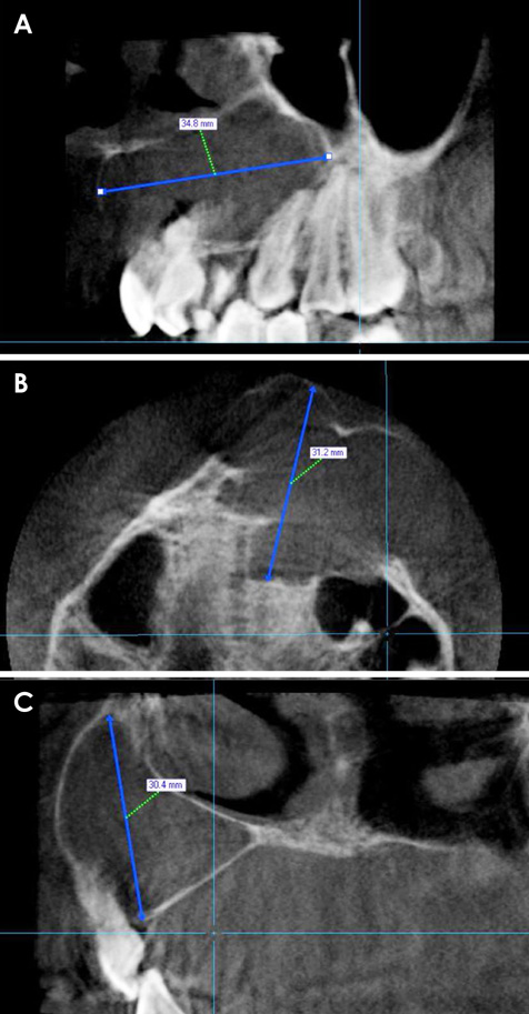
Fig. 1 Cone-beam computed tomographic images of a dentigerous cyst before surgery. A. The mesiodistal dimension was measured on the sagittal image. B. The anteroposterior dimension was measured on the axial image. C. The superoinferior dimension was measured on the coronal image.
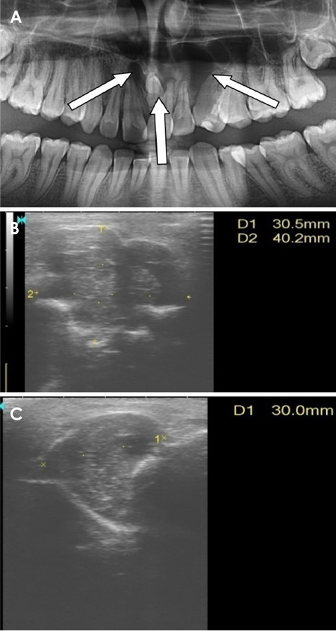
Fig. 2 A. A panoramic image shows the same dentigerous cyst around the mesiodens in the anterior maxilla. B. The mesiodistal and anteroposterior dimensions were measured on the ultrasound longitudinal scan. C. The superoinferior dimension of the same lesion before surgery was measured on the ultrasound transverse scan.
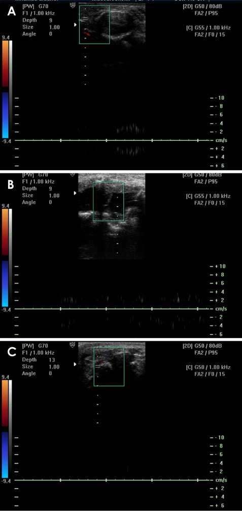
Fig. 3 A. Blood-flow velocity around the same lesion before surgery. B. Blood-flow velocity around the surgical site at 1 week after surgery in the same case. C. Blood-flow velocity around the surgical site at 6 months after surgery in the same case.

Fig. 4 Cone-beam computed tomographic images at 6 months after surgery from the same case. A. The mesiodistal dimension was measured on the sagittal image. B. The anteroposterior dimension was measured on the axial image. C. The superoinferior dimension was measured on the coronal image.

Fig. 5 Ultrasonographic images at 6 months after surgery from the same case. A. The mesiodistal and anteroposterior dimensions were measured on the longitudinal scan. Arrows show new bone formation. B. The superoinferior dimension was measured on the transverse scan.
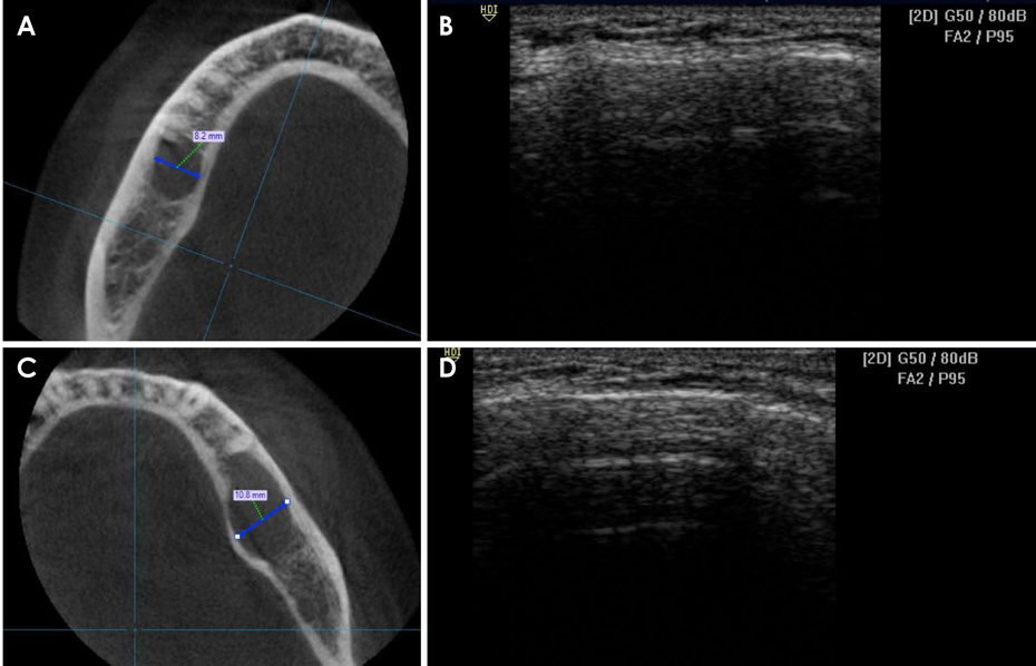
Fig. 6 A cone-beam computed tomographic (CBCT) image (A) and an ultrasonographic (US) image (B) show the first of 2 cases in which US waves could not penetrate the thick buccal bone plate. CBCT (C) and US (D) images show the second of 2 cases in which US waves could not penetrate the thick buccal bone plate.
Reference
-
1. Weber AL. Imaging of cysts and odontogenic tumors of the jaw. Definition and classification. Radiol Clin North Am. 1993; 31:101–120.2. Underhill TE, Katz JO, Pope TL Jr, Dunlap CL. Radiologic findings of diseases involving the maxilla and mandible. AJR Am J Roentgenol. 1992; 159:345–350.
Article3. Mast HL, Haller JO, Solomon M. Benign lesions of the mandibular and maxillary region in children: characterization by CT and MRI. Comput Med Imaging Graph. 1992; 16:1–9.
Article4. Abrahams JJ, Oliverio PJ. Odontogenic cysts: improved imaging with a dental CT software program. AJNR Am J Neuroradiol. 1993; 14:367–374.5. Araki M, Matsumoto N, Matsumoto K, Ohnishi M, Honda K, Komiyama K. Asymptomatic radiopaque lesions of the jaws: a radiographic study using cone-beam computed tomography. J Oral Sci. 2011; 53:439–444.
Article6. Dula K, Mini R, van der Stelt PF, Lambrecht JT, Schneeberger P, Buser D. Hypothetical mortality risk associated with spiral computed tomography of the maxilla and mandible. Eur J Oral Sci. 1996; 104:503–510.
Article7. Whaites E. Essentials of dental radiography and radiology. 4th ed. London: Churchill Livingstone;2007.8. Rajendran N, Sundaresan B. Efficacy of ultrasound and color power Doppler as a monitoring tool in the healing of endodontic periapical lesions. J Endod. 2007; 33:181–186.
Article9. Koller H, Kolb K, Zenner J, Reynolds J, Dvorak M, Acosta F, et al. Study on accuracy and interobserver reliability of the assessment of odontoid fracture union using plain radiographs or CT scans. Eur Spine J. 2009; 18:1659–1668.
Article10. Cotti E, Campisi G, Ambu R, Dettori C. Ultrasound real-time imaging in the differential diagnosis of periapical lesions. Int Endod J. 2003; 36:556–556.
Article11. Lauria L, Curi MM, Chammas MC, Pinto DS, Torloni H. Ultrasonography evaluation of bone lesions of the jaw. Oral Surg Oral Med Oral Pathol Oral Radiol Endod. 1996; 82:351–357.12. Tsiolis FI, Needleman IG, Griffiths GS. Periodontal ultrasonography. J Clin Periodontol. 2003; 30:849–854.
Article13. Imbeau J. Introduction to through-transmission alveolar ultrasonography (TAU) in dental medicine. Cranio. 2005; 23:100–112.
Article14. Tikku AP, Kumar S, Loomba K, Chandra A, Verma P, Aggarwal R. Use of ultrasound, color Doppler imaging and radiography to monitor periapical healing after endodontic surgery. J Oral Sci. 2010; 52:411–416.
Article15. Maity I, Kumari A, Shukla AK, Usha H, Naveen D. Monitoring of healing by ultrasound with color power Doppler after root canal treatment of maxillary anterior teeth with periapical lesions. J Conserv Dent. 2011; 14:252–257.
Article16. Lascala CA, Panella J, Marques MM. Analysis of the accuracy of linear measurements obtained by cone beam computed tomography (CBCT-NewTom). Dentomaxillofac Radiol. 2004; 33:291–294.
Article17. Kamburoğlu K, Kolsuz E, Kurt H, Kiliç C, Özen T, Paksoy CS. Accuracy of CBCT measurements of a human skull. J Digit Imaging. 2011; 24:787–793.
Article18. Ganguly R, Ruprecht A, Vincent S, Hellstein J, Timmons S, Qian F. Accuracy of linear measurement in the Galileos cone beam computed tomography under simulated clinical conditions. Dentomaxillofac Radiol. 2011; 40:299–305.
Article19. Stratemann SA, Huang JC, Maki K, Miller AJ, Hatcher DC. Comparison of cone beam computed tomography imaging with physical measures. Dentomaxillofac Radiol. 2008; 37:80–93.
Article20. Marotti J, Heger S, Tinschert J, Tortamano P, Chuembou F, Radermacher K, et al. Recent advances of ultrasound imaging in dentistry - a review of the literature. Oral Surg Oral Med Oral Pathol Oral Radiol. 2013; 115:819–832.
Article21. Mehdizadeh M, Movahedian B, Babasafari M, Mohammadi P. Comparison ultrasound, indirect digital panoramic radiography in differentional radiolucent mandible lesions. Res J Biol Sci. 2009; 4:1169–1170.22. Sumer AP, Danaci M, Ozen Sandikci E, Sumer M, Celenk P. Ultrasonography and Doppler ultrasonography in the evaluation of intraosseous lesions of the jaws. Dentomaxillofac Radiol. 2009; 38:23–27.
Article23. Goel S, Nagendrareddy SG, Raju MS, Krishnojirao DR, Rastogi R, Mohan RP, et al. Ultrasonography with color Doppler and power Doppler in the diagnosis of periapical lesions. Indian J Radiol Imaging. 2011; 21:279–283.
Article24. Gundappa M, Ng SY, Whaites EJ. Comparison of ultrasound, digital and conventional radiography in differentiating periapical lesions. Dentomaxillofac Radiol. 2006; 35:326–333.
Article25. Shahidi S, Shakibafard A, Zamiri B, Mokhtare MR, Houshyar M, Mahdian S. The feasibility of ultrasonography in defining the size of jaw osseous lesions. J Dent (Shiraz). 2015; 16:335–340.26. Bayrakdar IS, Yilmaz AB, Caglayan F, Ertas U, Gundogdu C, Gumussoy I. Cone beam computed tomography and ultrasonography imaging of benign intraosseous jaw lesion: a prospective radiopathological study. Clin Oral Investig. 2018; 22:1531–1539.
Article27. Moed BR, Subramanian S, van Holsbeeck M, Watson JT, Cramer KE, Karges DE, et al. Ultrasound for the early diagnosis of tibial fracture healing after static interlocked nailing without reaming: clinical results. J Orthop Trauma. 1998; 12:206–213.
Article28. Aggarwal V, Singla M. Use of computed tomography scans and ultrasound in differential diagnosis and evaluation of nonsurgical management of periapical lesions. Oral Surg Oral Med Oral Pathol Oral Radiol Endod. 2010; 109:917–923.
Article
- Full Text Links
-
- Actions
-
Cited
- CITED
-
- Close
- Share
-
- Similar articles
-
- Dental implant treatment after healing of bisphosphonate-related osteonecrosis of the jaw (BRONJ) in the same region: a case report
- Radiologic assessment of bone healing by fractal analysis after the treatment of jaw bone cyst by decompression
- Evaluation of ultrasonography as a diagnostic tool in the management of periapical cysts and granulomas: A clinical study
- Long-Term Efficacy and Safety of Denosumab: Insights beyond 10 Years of Use
- Diagnosis of Cystic Hepatic Lesions on Ultrasonography
