J Korean Ophthalmol Soc.
2018 Sep;59(9):876-880. 10.3341/jkos.2018.59.9.876.
Bilateral Frosted Branch Angiitis in Kikuchi-Fujimoto Disease
- Affiliations
-
- 1Department of Ophthalmology, Chosun University School of Medicine, Gwangju, Korea. s20age@hanmail.net
- 2Gangjin Medical Center, Sinan, Korea.
- KMID: 2420338
- DOI: http://doi.org/10.3341/jkos.2018.59.9.876
Abstract
- PURPOSE
A case of frosted branch angiitis in Kikuchi-Fujimoto disease is reported.
CASE SUMMARY
A 33-year-old male complained of a sudden decrease in visual acuity that developed in both eyes 5 days prior. He suffered from a headache, chills, myalgia, and flank pain 1 week before. The initial best-corrected visual acuity (BCVA) was 0.1 in the right eye and 0.2 in the left eye. On slit lamp examination, no inflammatory finding was observed in the anterior chamber and vitreous body of both eyes. On fundus examination, a diffuse vascular sheathing-like frosted branch was found in the retinal vessels, and retinal hemorrhage was observed. Fluorescein angiography showed staining and leakage of dye along the vascular sheathing. Serological findings were negative, showing no evidence of an autoimmune disease or viral infection. Neck ultrasonography revealed non-tender left cervical lymph node enlargement >1 cm in diameter. Ultrasound-guided fine needle aspiration cytology showed findings compatible with Kikuchi-Fujimoto disease, including necrotic changes and pronounced karyorrhexis, plus histiocyte and lymphocyte infiltration without neutrophils. We started systemic steroid therapy. One month after treatment, the BCVA of both eyes improved to 1.0.
CONCLUSIONS
In patients with frosted branch angiitis, systemic disease such as Kikuchi-Fujimoto disease should be considered.
MeSH Terms
-
Adult
Anterior Chamber
Autoimmune Diseases
Biopsy, Fine-Needle
Chills
Flank Pain
Fluorescein Angiography
Headache
Histiocytes
Histiocytic Necrotizing Lymphadenitis*
Humans
Lymph Nodes
Lymphocytes
Male
Myalgia
Neck
Neutrophils
Retinal Hemorrhage
Retinal Vessels
Slit Lamp
Ultrasonography
Vasculitis*
Visual Acuity
Vitreous Body
Figure
-
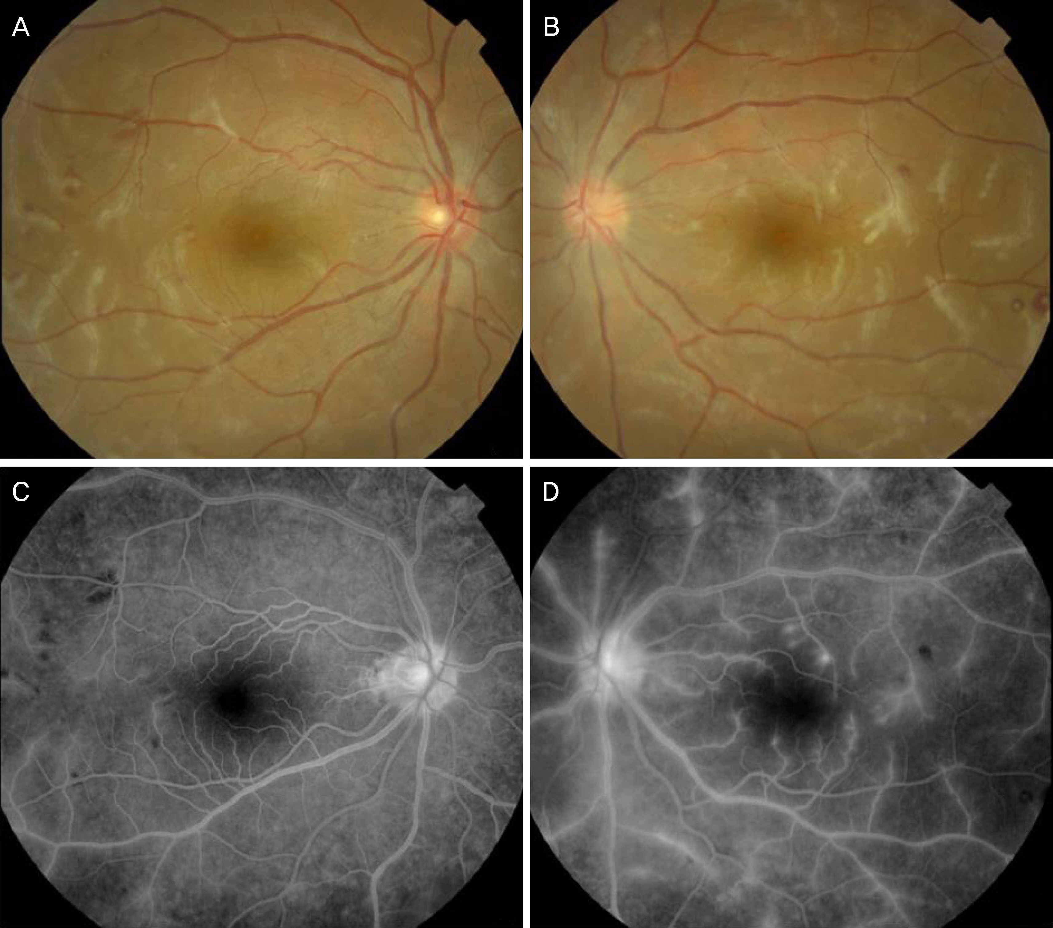
Figure 1. Fundus exam at the initial visit. Fundus photograph shows sheathing of retinal vessels and multiple retinal hemorrhages in the right (A) and the left (B) eye. Fundus fluorescein angiography at the initial examination shows dye leakages from the sheathed retinal vessels with normal blood flow in the right (C) and the left (D) eye.
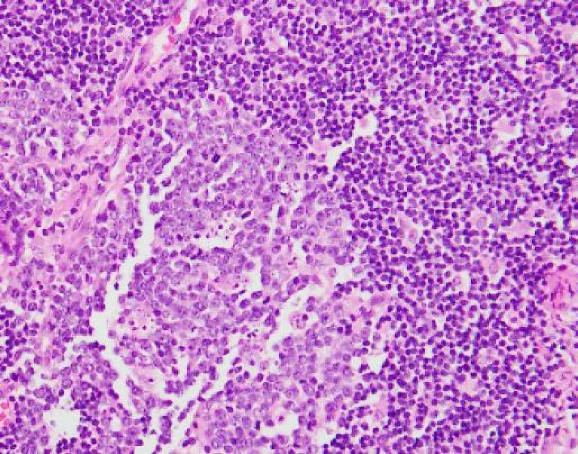
Figure 2. Histopathological finding of the cervical lymph node. Numerous phagocytic histiocytes and prominent karyorrhexis are identified (hematoxylin and eosin stain [H&E] stain, ×100).
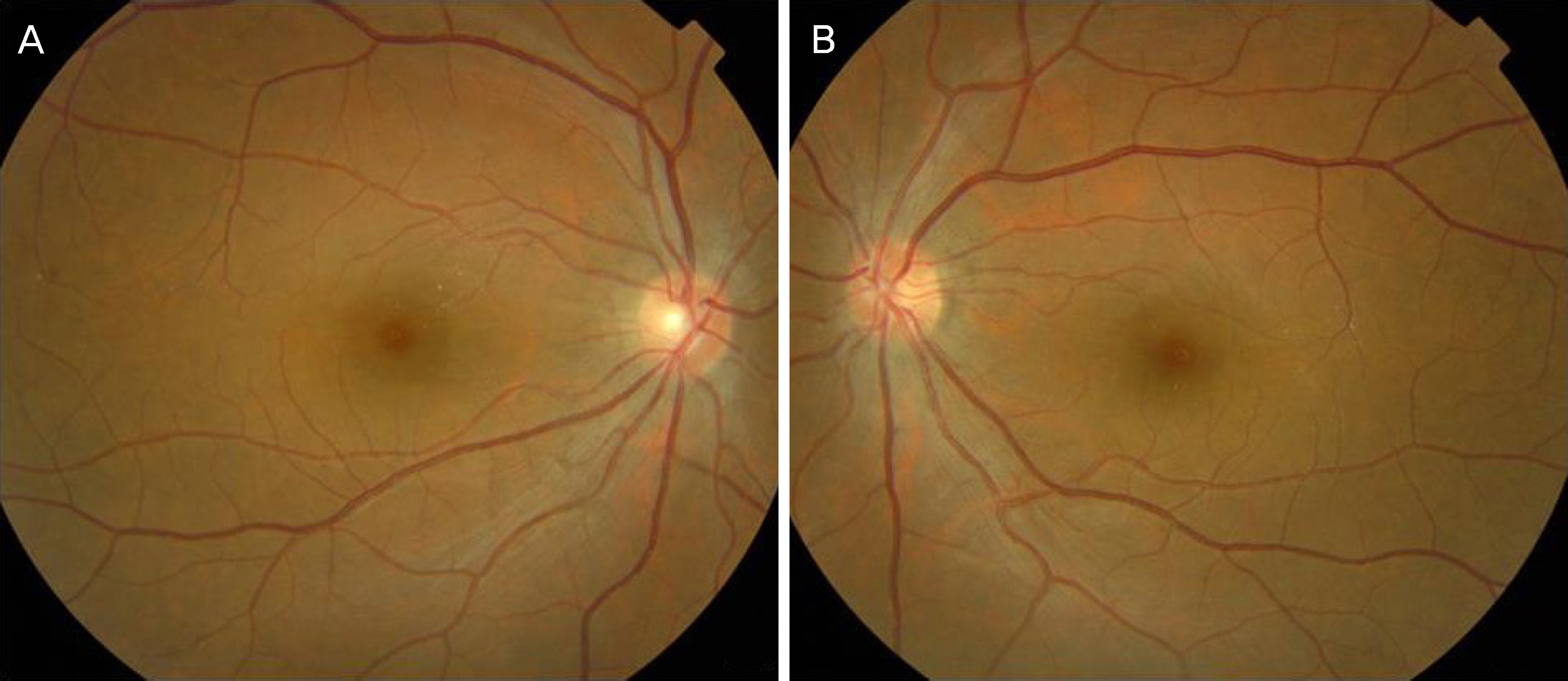
Figure 3. Fundus photographs at 1 month after systemic steroid therapy. Fundus photograph shows almost normal without any sheathing of retinal vessels in the right (A) and the left (B) eye.
Reference
-
References
1. Norris AH, Krasinskas AM, Salhany KE, Gluckmman SJ. KikuchiFujimoto disease: a benign cause of fever and lymphadenopathy. Am J Med. 1996; 101:401–5.
Article2. Rocher F, Pelosse B, Momtchilova M, Laroche L. Kikuchi's abdominal and ocular manifestation. J Fr Ophtalmal. 2006; 29:932–6.3. Ito Y. Frosted branch angiitis in a child. Jpn J Clin Ophthalmol. 1976; 30:797–803.4. Bosch X, Guilabert A, Miquel R, et al. Enigmatic Kikuchi-Fujimoto disease: a comprehensive review. Am J Clin Pathol. 2004; 122:141–52.5. Famularo G, Giustiniani MC, Marasco A, et al. De Simone C. Kikuchi Fujimoto lymphadenitis: case report and literature review. AM J Hematol. 2003; 74:60–3.6. Cheng CY, Sheng WH, Lo YC, et al. Clinical presentations, abdominal results and outcomes of patients with Kikuchi's disease: abdominal on the association between recurrent Kikuchi's disease and autoimmune diseases. J Microbiol Immunol Infect. 2010; 43:366–71.7. Kim SH, Kim SJ, Chung H, et al. Bilateral anterior uveitis as an abdominal manifestation of Kikuchi-Fujimoto disease. Rheumatology (Oxford). 2004; 43:1056–7.8. Taguri AH, Mcllwaine GG. Bilateral panuveitis: a possible abdominal with Kikuchi–Fujimoto disease. Am J Ophthalmol. 2001; 132:419–21.9. Zou W, Wen F. Bilateral occlusive retinal vasculitis in Kikuchi-Fujumoto disease. Clin Exp Ophthalmol. 2007; 35:875–7.10. Galor A, Georgy M, Leder HA, et al. Papillary conjunctivitis abdominal with Kikuchi disease. Cornea. 2008; 27:944–6.11. Biswas J, Fogla R, Madhavan HN. Bilateral frosted branchangiitis in an 8-year-old indian girl. Retina. 1996; 16:444–5.12. Masuda K, Ueno M, Watanabe I. A case of frosted branch angiitis with yellowish-white placoid lesions: fluorescein and indocyanine green angiography findings. Jpn J Ophthalmol. 1998; 42:484–9.
Article13. Duker JS, Brown GC, Brooks L. Retinal vasculitis in Crohn's disease. Am J Ophthalmol. 1987; 103:664–8.
Article14. Dorfman RF, Berry GJ. Kikuchi's histiocytic necrotizing lympha-denitis: an analysis of 108 cases with emphasis of differential diagnosis. Semin Diagn Pathol. 1988; 5:329–45.15. Imamura M, Ueno H, Matsuura A, et al. An ultrastructural study of subacute necrotizing lymphadenitis. Am J Pathol. 1982; 107:292–9.
- Full Text Links
-
- Actions
-
Cited
- CITED
-
- Close
- Share
-
- Similar articles
-
- Frosted Branch Angiitis Associated with COVID-19-related Multisystem Inflammatory Syndrome in Children
- Bilateral Frosted Branch Angiitis in Anti-phospholipid Antibody Syndrome
- Frosted Branch Angiitis as Ocular Manifestation of Behcet's Disease: Unusual Case Report and Literature Review
- Frosted Branch Angiitis
- A Case of Frosted Branch Angiitis
