Ann Lab Med.
2019 Jan;39(1):91-95. 10.3343/alm.2019.39.1.91.
Dose Estimation Curves Following In Vitro X-ray Irradiation Using Blood From Four Healthy Korean Individuals
- Affiliations
-
- 1Department of Laboratory Medicine and Genetics, Soonchunhyang University Bucheon Hospital, Soonchunhyang University College of Medicine, Bucheon, Korea. cecilia@schmc.ac.kr, shinhb@schmc.ac.kr
- 2Department of Laboratory Medicine, Korea Cancer Center Hospital, Korea Institute of Radiological and Medical Sciences, Seoul, Korea.
- 3Department of Radiation Oncology, Soonchunhyang University Bucheon Hospital, Soonchunhyang University College of Medicine, Bucheon, Korea.
- KMID: 2420277
- DOI: http://doi.org/10.3343/alm.2019.39.1.91
Abstract
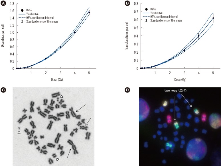
- Cytogenetic dosimetry is useful for evaluating the absorbed dose of ionizing radiation based on analysis of radiation-induced chromosomal aberrations. We created two types of in vitro dose-response calibration curves for dicentric chromosomes (DC) and translocations (TR) induced by X-ray irradiation, using an electron linear accelerator, which is the most frequently used medical device in radiotherapy. We irradiated samples from four healthy Korean individuals and compared the resultant curves between individuals. Aberration yields were studied in a total of 31,800 and 31,725 metaphases for DC and TR, respectively, obtained from 11 X-ray irradiation dose-points (0, 0.05, 0.1, 0.25, 0.5, 0.75, 1, 2, 3, 4, and 5 Gy). The dose-response relationship followed a linear-quadratic equation, Y=C+αD+βD², with the coefficients C=0.0011 for DC and 0.0015 for TR, α=0.0119 for DC and 0.0048 for TR, and β=0.0617 for DC and 0.0237 for TR. Correlation coefficients between irradiation doses and chromosomal aberrations were 0.971 for DC and 0.6 for TR, indicating a very strong and a moderate correlation, respectively. This is the first study implementing cytogenetic dosimetry following exposure to ionizing X-radiation.
Keyword
MeSH Terms
Figure
Reference
-
1. Ricoul M, Gnana-Sekaran T, Piqueret-Stephan L, Sabatier L. Cytogenetics for biological dosimetry. Methods Mol Biol. 2017; 1541:189–208. PMID: 27910025.2. International Atomic Energy Agency. Cytogenetic dosimetry: applications in preparedness for and response to radiation emergencies. Vienna: International Atomic Energy Agency Publishing;2011. p. 45–202.3. Pinto MM, Santos NF, Amaral A. Current status of biodosimetry based on standard cytogenetic methods. Radiat Environ Biophys. 2010; 49:567–581. PMID: 20617329.4. Cho MS, Lee JK, Bae KS, Han EA, Jang SJ, Ha WH, et al. Retrospective biodosimetry using translocation frequency in a stable cell of occupationally exposed to ionizing radiation. J Radiat Res. 2015; 56:709–716. PMID: 25922373.5. Kang CM, Yun HJ, Kim H, Kim CS. Strong correlation among three biodosimetry techniques following exposures to ionizing radiation. Genome Integr. 2016; 7:11. PMID: 28217287.6. Ministry of Health and Welfare. Accessed on July 2018. http://www.mohw.go.kr/react/jb/sjb0406ls.jsp?PAR_MENU_ID=03&MENU_ID=030406.7. Romm H, Oestreicher U, Kulka U. Cytogenetic damage analysed by the dicentric assay. Ann Ist Super Sanita. 2009; 45:251–259. PMID: 19861729.8. Lee JK. Practical applications of cytogenetic biodosimetry in radiological emergencies. Korean J Hematol. 2011; 46:62–64. PMID: 21747874.9. Lemos-Pinto MM, Cadena M, Santos N, Fernandes TS, Borges E, Amaral A. A dose-response curve for biodosimetry from a 6 MV electron linear accelerator. Braz J Med Biol Res. 2015; 48:908–914. PMID: 26445334.10. Healy BJ, van der Merwe D, Christaki KE, Meghzifene A. Cobalt-60 machines and medical linear accelerators: competing technologies for external beam radiotherapy. Clin Oncol (R Coll Radiol). 2017; 29:110–115. PMID: 27908503.11. Mukaka MM. Statistics corner: A guide to appropriate use of correlation coefficient in medical research. Malawi Med J. 2012; 24:69–71. PMID: 23638278.12. Suto Y, Akiyama M, Noda T, Hirai M. Construction of a cytogenetic dose-response curve for low-dose range gamma-irradiation in human peripheral blood lymphocytes using three-color FISH. Mutat Res Genet Toxicol Environ Mutagen. 2015; 794:32–38. PMID: 26653981.13. Abe Y, Yoshida MA, Fujioka K, Kurosu Y, Ujiie R, Yanagi A, et al. Dose-response curves for analyzing of dicentric chromosomes and chromosome translocations following doses of 1000 mGy or less, based on irradiated peripheral blood samples from five healthy individuals. J Radiat Res. 2018; 59:35–42. PMID: 29040682.14. Wilkins RC, Romm H, Kao TC, Awa AA, Yoshida MA, Livingston GK, et al. Interlaboratory comparison of the dicentric chromosome assay for radiation biodosimetry in mass casualty events. Radiat Res. 2008; 169:551–560. PMID: 18439045.15. Ryu TH, Kim JH, Kim JK. Chromosomal aberrations in human peripheral blood lymphocytes after exposure to ionizing radiation. Genome Integr. 2016; 7:5. PMID: 28217281.
- Full Text Links
- Actions
-
Cited
- CITED
-
- Close
- Share
- Similar articles
-
- Radiation-Induced Apoptosis of Lymphocytes in Peripheral Blood
- A Stereotactic Device for Gamma Knife Irradiation to Cell Lines
- Effect of whole body x-irradiation on the NP-SH level of blood in rabbits
- Effect of Strontium 90 Beta-Irradiation, In Vitro on the Distensibility of the Cornea and Sclera of the Rabbit
- Dose-Response Curves of Mouse Jejunal Crypt Cells by Multifrationated Irrdiation


