Osteotomy around the Knee: Indication and Preoperative Planning
- Affiliations
-
- 1Department of Orthopedic Surgery, Yeungnam University Hospital, Daegu, Korea. aestro-jin@hanmail.net
- KMID: 2419463
- DOI: http://doi.org/10.4055/jkoa.2018.53.4.283
Abstract
- Osteotomy around the knee is a widely considered surgical procedure for osteoarthritis with lower extremity malalignment. High tibial osteotomy (HTO) is performed for varus deformity, while distal femur osteotomy (DFO) is performed for valgus deformity. However, if the correction is insufficient, double osteotomy can also be considered. This report included the basic principles and current concepts of patient selection and preoperative planning in osteotomy around the knee.
Keyword
MeSH Terms
Figure
-
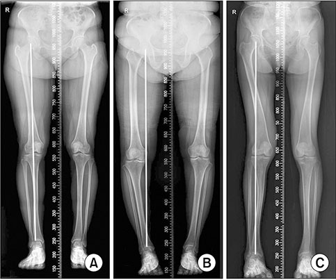
Figure 1 (A) Normal axis alignment. (B) Varus axis alignment. (C) Valgus axis alignment.
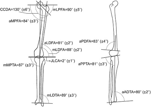
Figure 2 Normal physiologic axes and angles of the lower extremities: The physiologic axes and angles have an abnormal value in the malalignment of the lower extremities. CCDA, centrum column diaphyseal angle; aMPFA, anatomical medial proximal femoral angle; mLPFA, mechanical lateral proximal femoral angle; aLDFA, anatomical lateral distal femoral angle; mLDFA, mechanical lateral distal femoral angle; JLCA, joint line congruency angle; mMPTA, mechanical medial proximal tibial angle; mLDTA, mechanical lateral distal tibial angle; aPDFA, anatomical posterior distal femoral angle; aPPTA, anatomical posterior proximal tibial angle; aADTA, anatomical anterior distal tibial angle.
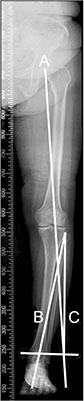
Figure 3 Bilateral weight bearing anteroposterior whole lower limb x-ray in full extension for planning the Miniaci method. Line A represents the planned weight bearing line for the postoperative correction extending from the center of the hip to about 60% to 70% of the tibial plateau width past the ankle. Line B connects the osteotomy hinge point with the center of the ankle. Line C connects the osteotomy hinge point with the arc intersection of Line A. The angle formed by Lines B and C is the planned correction angle.
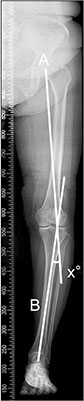
Figure 4 Bilateral weight bearing anteroposterior whole lower limb x-ray in full extension for planning the Dugdale method. Line A is drawn from the center of the femoral head to 62.5% of the tibial width. Line B is drawn from the center of the tibiotalar joint to the 62.5% coordinate. The angle formed by these two lines is the correction angle.
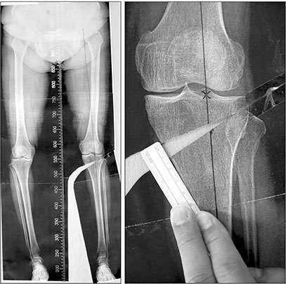
Figure 5 Weight bearing scanography. A template was cut through the osteotomy site, and the tibia was rotated until the weight-bearing line passed through the 62% to 65% coordinate.
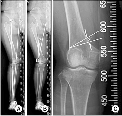
Figure 6 Bilateral weight bearing anteroposterior whole lower limb x-ray in full extension to plan the medial close-wedge distal femoral osteotomy. (A) The present mechanical axis is drawn from A, which is the center of the femoral head, to B, which is the center of the ankle joint. Line B–C is of equal length as Line A–B and passes the knee, just medial to the medial eminence, representing the desired postoperative mechanical axis. (B) The hinge point of the osteotomy D is marked just proximal to the upper border of the lateral condyle and 0.5–1.0 cm within the lateral cortex. The angle of correction (α) is defined by Line A–D between the present femoral head center and the hinge point and Line C–D connecting the new femoral head center position and the hinge point. (C) Correction angle α is projected at the distal femur using two oblique, down-sloping lines of equal length converging at the hinge point. The distance measured between those two lines at the level of the medial cortex represents the osteotomy wedge base length, which is to be removed during surgery.
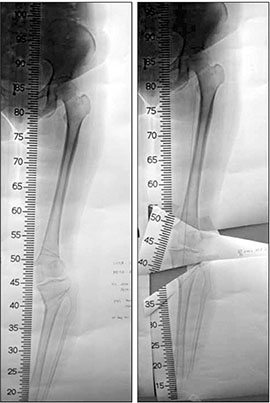
Figure 7 Double level osteotomy. A template was cut through each osteotomy site, and the femur and tibia were rotated until the weight-bearing line passed through the 62% to 65% coordinate (Courtesy of Professor Dr. Seung-Beom Han, Department of Orthopaedic Surgery, Korea University Anam Hospital, Korea University College of Medicine, Seoul, Korea)
Reference
-
1. Nakagawa Y, Mukai S, Yabumoto H, Tarumi E, Nakamura T. Cartilage degeneration and alignment in severe varus knee osteoarthritis. Cartilage. 2015; 6:208–215.
Article2. Aglietti P, Rinonapoli E, Stringa G, Taviani A. Tibial osteotomy for the varus osteoarthritic knee. Clin Orthop Relat Res. 1983; 176:239–251.
Article3. Coventry MB. Proximal tibial varus osteotomy for osteoarthritis of the lateral compartment of the knee. J Bone Joint Surg Am. 1987; 69:32–38.
Article4. Insall JN, Joseph DM, Msika C. High tibial osteotomy for varus gonarthrosis. A long-term follow-up study. J Bone Joint Surg Am. 1984; 66:1040–1048.
Article5. McDermott AG, Finklestein JA, Farine I, Boynton EL, MacIntosh DL, Gross A. Distal femoral varus osteotomy for valgus deformity of the knee. J Bone Joint Surg Am. 1988; 70:110–116.
Article6. Morrey BF. Upper tibial osteotomy for secondary osteoarthritis of the knee. J Bone Joint Surg Br. 1989; 71:554–559.
Article7. Morrey BF, Edgerton BC. Distal femoral osteotomy for lateral gonarthrosis. Instr Course Lect. 1992; 41:77–85.8. Rinonapoli E, Mancini GB, Corvaglia A, Musiello S. Tibial osteotomy for varus gonarthrosis. A 10- to 21-year followup study. Clin Orthop Relat Res. 1998; 353:185–193.9. Bonasia DE, Dettoni F, Sito G, et al. Medial opening wedge high tibial osteotomy for medial compartment overload/arthritis in the varus knee: prognostic factors. Am J Sports Med. 2014; 42:690–698.10. Coventry MB, Ilstrup DM, Wallrichs SL. Proximal tibial osteotomy. A critical long-term study of eighty-seven cases. J Bone Joint Surg Am. 1993; 75:196–201.
Article11. Hunter DJ, Sharma L, Skaife T. Alignment and osteoarthritis of the knee. J Bone Joint Surg Am. 2009; 91:Suppl 1. 85–89.
Article12. Amis AA. Biomechanics of high tibial osteotomy. Knee Surg Sports Traumatol Arthrosc. 2013; 21:197–205.
Article13. Leonardi F, Rivera F, Zorzan A, Ali SM. Bilateral double osteotomy in severe torsional malalignment syndrome: 16 years follow-up. J Orthop Traumatol. 2014; 15:131–136.14. Yoo JD, Kim NK. Distal femoral varization osteotomy. J Korean Orthop Assoc. 2014; 49:118–125.
Article15. Hsu RW, Himeno S, Coventry MB, Chao EY. Normal axial alignment of the lower extremity and load-bearing distribution at the knee. Clin Orthop Relat Res. 1990; 255:215–227.
Article16. Moreland JR, Bassett LW, Hanker GJ. Radiographic analysis of the axial alignment of the lower extremity. J Bone Joint Surg Am. 1987; 69:745–749.
Article17. Tetsworth K, Paley D. Malalignment and degenerative arthropathy. Orthop Clin North Am. 1994; 25:367–377.
Article18. Fujisawa Y, Masuhara K, Shiomi S. The effect of high tibial osteotomy on osteoarthritis of the knee. An arthroscopic study of 54 knee joints. Orthop Clin North Am. 1979; 10:585–608.19. Moore J, Mychaltchouk L, Lavoie F. Applicability of a modified angular correction measurement method for open-wedge high tibial osteotomy. Knee Surg Sports Traumatol Arthrosc. 2017; 25:846–852.
Article20. Pape D, Rupp S. Preoperative planning for high tibial osteotomies. Oper Tech Orthop. 2007; 17:2–11.
Article21. Brown GA, Amendola A. Radiographic evaluation and preoperative planning for high tibial osteotomies. Oper Tech Sports Med. 2012; 20:93–102.
Article22. Brinkman JM, Lobenhoffer P, Agneskirchner JD, Staubli AE, Wymenga AB, van Heerwaarden RJ. Osteotomies around the knee: patient selection, stability of fixation and bone healing in high tibial osteotomies. J Bone Joint Surg Br. 2008; 90:1548–1557.23. Flecher X, Parratte S, Aubaniac JM, Argenson JN. A 12-28-year followup study of closing wedge high tibial osteotomy. Clin Orthop Relat Res. 2006; 452:91–96.
Article24. Khoshbin A, Sheth U, Ogilvie-Harris D, et al. The effect of patient, provider and surgical factors on survivorship of high tibial osteotomy to total knee arthroplasty: a population-based study. Knee Surg Sports Traumatol Arthrosc. 2017; 25:887–894.
Article25. Naudie D, Bourne RB, Rorabeck CH, Bourne TJ. The Install Award. Survivorship of the high tibial valgus osteotomy. A 10- to -22-year followup study. Clin Orthop Relat Res. 1999; 367:18–27.26. Trieb K, Grohs J, Hanslik-Schnabel B, Stulnig T, Panotopoulos J, Wanivenhaus A. Age predicts outcome of high-tibial osteotomy. Knee Surg Sports Traumatol Arthrosc. 2006; 14:149–152.
Article27. Goshima K, Sawaguchi T, Sakagoshi D, Shigemoto K, Hatsuchi Y, Akahane M. Age does not affect the clinical and radiological outcomes after open-wedge high tibial osteotomy. Knee Surg Sports Traumatol Arthrosc. 2017; 25:918–923.
Article28. Odenbring S, Egund N, Knutson K, Lindstrand A, Larsen ST. Revision after osteotomy for gonarthrosis. A 10-19-year follow-up of 314 cases. Acta Orthop Scand. 1990; 61:128–130.
Article29. van Raaij T, Reijman M, Brouwer RW, Jakma TS, Verhaar JN. Survival of closing-wedge high tibial osteotomy: good outcome in men with low-grade osteoarthritis after 10-16 years. Acta Orthop. 2008; 79:230–234.30. Shin YS, Lee DH, Lee SH, Kim MJ, Han SB. Basic principles and current trends of medial opening-wedge high tibial osteotomy. J Korean Orthop Assoc. 2014; 49:85–94.
Article31. Nwachukwu BU, McCormick FM, Schairer WW, Frank RM, Provencher MT, Roche MW. Unicompartmental knee arthroplasty versus high tibial osteotomy: United States practice patterns for the surgical treatment of unicompartmental arthritis. J Arthroplasty. 2014; 29:1586–1589.
Article32. Fu D, Li G, Chen K, Zhao Y, Hua Y, Cai Z. Comparison of high tibial osteotomy and unicompartmental knee arthroplasty in the treatment of unicompartmental osteoarthritis: a meta-analysis. J Arthroplasty. 2013; 28:759–765.33. W-Dahl A, Toksvig-Larsen S, Lindstrand A. Ten-year results of physical activity after high tibial osteotomy in patients with knee osteoarthritis. Knee Surg Sports Traumatol Arthrosc. 2017; 25:902–909.
Article34. W-Dahl A, Robertsson O, Lidgren L. Surgery for knee osteoarthritis in younger patients. Acta Orthop. 2010; 81:161–164.
Article35. Börjesson M, Weidenhielm L, Mattsson E, Olsson E. Gait and clinical measurements in patients with knee osteoarthritis after surgery: a prospective 5-year follow-up study. Knee. 2005; 12:121–127.
Article36. Stukenborg-Colsman C, Wirth CJ, Lazovic D, Wefer A. High tibial osteotomy versus unicompartmental joint replacement in unicompartmental knee joint osteoarthritis: 7-10-year follow-up prospective randomised study. Knee. 2001; 8:187–194.37. Bonnin M, Chambat P. Current status of valgus angle, tibial head closing wedge osteotomy in media gonarthrosis. Orthopade. 2004; 33:135–142.38. Kumagai K, Akamatsu Y, Kobayashi H, Kusayama Y, Koshino T, Saito T. Factors affecting cartilage repair after medial opening-wedge high tibial osteotomy. Knee Surg Sports Traumatol Arthrosc. 2017; 25:779–784.
Article39. Pape D, Seil R, Adam F, Rupp S, Kohn D, Lobenhoffer P. Imaging and preoperative planning of osteotomy of tibial head osteotomy. Orthopade. 2004; 33:122–134.40. OAhlbäck S. Osteoarthrosis of the knee. A radiographic investigation. Acta Radiol Diagn (Stockh). 1968; Suppl 277. 7–72.41. Murphy SB. Tibial osteotomy for genu varum. Indications, preoperative planning, and technique. Orthop Clin North Am. 1994; 25:477–482.42. Berman AT, Bosacco SJ, Kirshner S, Avolio A Jr. Factors influencing long-term results in high tibial osteotomy. Clin Orthop Relat Res. 1991; 272:192–198.
Article43. Akizuki S, Shibakawa A, Takizawa T, Yamazaki I, Horiuchi H. The long-term outcome of high tibial osteotomy: a ten- to 20-year follow-up. J Bone Joint Surg Br. 2008; 90:592–596.44. Nha KW, Kim HJ, Ahn HS, Lee DH. Change in posterior tibial slope after open-wedge and closed-wedge high tibial osteotomy: a meta-analysis. Am J Sports Med. 2016; 44:3006–3013.45. Naudie DD, Amendola A, Fowler PJ. Opening wedge high tibial osteotomy for symptomatic hyperextension-varus thrust. Am J Sports Med. 2004; 32:60–70.
Article46. Phisitkul P, Wolf BR, Amendola A. Role of high tibial and distal femoral osteotomies in the treatment of lateral-posterolateral and medial instabilities of the knee. Sports Med Arthrosc. 2006; 14:96–104.
Article47. Arthur A, LaPrade RF, Agel J. Proximal tibial opening wedge osteotomy as the initial treatment for chronic posterolateral corner deficiency in the varus knee: a prospective clinical study. Am J Sports Med. 2007; 35:1844–1850.48. Giagounidis EM, Sell S. High tibial osteotomy: factors influencing the duration of satisfactory function. Arch Orthop Trauma Surg. 1999; 119:445–449.
Article49. Lee YS, Lee BK, Lee SH, Park HG, Jun DS, Moon DH. Effect of foot rotation on the mechanical axis and correlation between knee and whole leg radiographs. Knee Surg Sports Traumatol Arthrosc. 2013; 21:2542–2547.
Article50. Lee YS, Lee BK, Kwon JH, et al. Serial assessment of weight-bearing lower extremity alignment radiographs after open-wedge high tibial osteotomy. Arthroscopy. 2014; 30:319–325.51. Sancheti P, Patil K, Gugale S, Shyam A. How can preoperative planning prevent occurrence of a painful total knee replacement?. In : Hirschmann MT, Becker R, editors. The unhappy total knee replacement. Cham: Springer;2015. p. 59–68.52. Phillips CL, Silver DA, Schranz PJ, Mandalia V. The measurement of patellar height: a review of the methods of imaging. J Bone Joint Surg Br. 2010; 92:1045–1053.53. Coventry MB. Upper tibial osteotomy for osteoarthritis. J Bone Joint Surg Am. 1985; 67:1136–1140.
Article54. Hernigou P, Medevielle D, Debeyre J, Goutallier D. Proximal tibial osteotomy for osteoarthritis with varus deformity. A ten to thirteen-year follow-up study. J Bone Joint Surg Am. 1987; 69:332–354.55. Miniaci A, Ballmer FT, Ballmer PM, Jakob RP. Proximal tibial osteotomy. A new fixation device. Clin Orthop Relat Res. 1989; 246:250–259.56. Dugdale TW, Noyes FR, Styer D. Preoperative planning for high tibial osteotomy. The effect of lateral tibiofemoral separation and tibiofemoral length. Clin Orthop Relat Res. 1992; 274:248–264.57. Jakob RP, Jacobi M. Closing wedge osteotomy of the tibial head in treatment of single compartment arthrosis. Orthopade. 2004; 33:143–152.58. Lee DH, Han SB, Oh KJ, et al. The weight-bearing scanogram technique provides better coronal limb alignment than the navigation technique in open high tibial osteotomy. Knee. 2014; 21:451–455.
Article59. Akamatsu Y, Mitsugi N, Mochida Y, et al. Navigated opening wedge high tibial osteotomy improves intraoperative correction angle compared with conventional method. Knee Surg Sports Traumatol Arthrosc. 2012; 20:586–593.
Article60. Reising K, Strohm PC, Hauschild O, et al. Computer-assisted navigation for the intraoperative assessment of lower limb alignment in high tibial osteotomy can avoid outliers compared with the conventional technique. Knee Surg Sports Traumatol Arthrosc. 2013; 21:181–188.
Article61. Elson DW, Petheram TG, Dawson MJ. High reliability in digital planning of medial opening wedge high tibial osteotomy, using Miniaci's method. Knee Surg Sports Traumatol Arthrosc. 2015; 23:2041–2048.
Article62. Lee DH, Nha KW, Park SJ, Han SB. Preoperative and postoperative comparisons of navigation and radiologic limb alignment measurements after high tibial osteotomy. Arthroscopy. 2012; 28:1842–1850.
Article63. Yoon SD, Zhang G, Kim HJ, Lee BJ, Kyung HS. Comparison of cable method and miniaci method using picture archiving and communication system in preoperative planning for open wedge high tibial osteotomy. Knee Surg Relat Res. 2016; 28:283–288.
Article64. Giffin JR, Vogrin TM, Zantop T, Woo SL, Harner CD. Effects of increasing tibial slope on the biomechanics of the knee. Am J Sports Med. 2004; 32:376–382.
Article65. Dean CS, Liechti DJ, Chahla J, Moatshe G, LaPrade RF. Clinical outcomes of high tibial osteotomy for knee instability: a systematic review. Orthop J Sports Med. 2016; 4:2325967116633419.66. Dejour H, Bonnin M. Tibial translation after anterior cruciate ligament rupture. Two radiological tests compared. J Bone Joint Surg Br. 1994; 76:745–749.
Article67. Chae DJ, Shetty GM, Lee DB, Choi HW, Han SB, Nha KW. Tibial slope and patellar height after opening wedge high tibia osteotomy using autologous tricortical iliac bone graft. Knee. 2008; 15:128–133.
Article68. Ogawa H, Matsumoto K, Ogawa T, Takeuchi K, Akiyama H. Effect of wedge insertion angle on posterior tibial slope in medial opening wedge high tibial osteotomy. Orthop J Sports Med. 2016; 4:2325967116630748.
Article69. Bae DK, Ko YW, Kim SJ, Baek JH, Song SJ. Computer-assisted navigation decreases the change in the tibial posterior slope angle after closed-wedge high tibial osteotomy. Knee Surg Sports Traumatol Arthrosc. 2016; 24:3433–3440.
Article70. Sharma L, Song J, Felson DT, Cahue S, Shamiyeh E, Dunlop DD. The role of knee alignment in disease progression and functional decline in knee osteoarthritis. JAMA. 2001; 286:188–195.
Article71. Stähelin T, Hardegger F, Ward JC. Supracondylar osteotomy of the femur with use of compression. Osteosynthesis with a malleable implant. J Bone Joint Surg Am. 2000; 82:712–722.72. Chahla J, Mitchell JJ, Liechti DJ, et al. Opening- and closing-wedge distal femoral osteotomy: a systematic review of outcomes for isolated lateral compartment osteoarthritis. Orthop J Sports Med. 2016; 4:2325967116649901.73. Hinterwimmer S, Rosenstiel N, Lenich A, Waldt S, Imhoff AB. Femoral osteotomy for patellofemoral instability. Unfallchirurg. 2012; 115:410–416.74. Niklasch M, Dreher T, Döderlein L, et al. Superior functional outcome after femoral derotation osteotomy according to gait analysis in cerebral palsy. Gait posture. 2015; 41:52–56.
Article75. Kazi HA, Abdel-Rahman W, Brady PA, Cameron JC. Meniscal allograft with or without osteotomy: a 15-year follow-up study. Knee Surg Sports Traumatol Arthrosc. 2015; 23:303–309.
Article76. Wang JW, Hsu CC. Distal femoral varus osteotomy for osteoarthritis of the knee. J Bone Joint Surg Am. 2005; 87:127–133.
Article77. Thein R, Bronak S, Thein R, Haviv B. Distal femoral osteotomy for valgus arthritic knees. J Orthop Sci. 2012; 17:745–749.
Article78. Backstein D, Morag G, Hanna S, Safir O, Gross A. Long-term follow-up of distal femoral varus osteotomy of the knee. J Arthroplasty. 2007; 22:S2–S6.
Article79. Finkelstein JA, Gross AE, Davis A. Varus osteotomy of the distal part of the femur. A survivorship analysis. J Bone Joint Surg Am. 1996; 78:1348–1352.
Article80. Franco V, Cipolla M, Gerullo G, Gianni E, Puddu G. Open wedge osteotomy of the distal femur in the valgus knee. Orthopade. 2004; 33:185–192.81. Mathews J, Cobb AG, Richardson S, Bentley G. Distal femoral osteotomy for lateral compartment osteoarthritis of the knee. Orthopedics. 1998; 21:437–440.
Article82. Paley D, Pfeil J. Principles of deformity correction around the knee. Orthopade. 2000; 29:18–38.
Article83. Benjamin A. Double osteotomy for the painful knee in rheumatoid arthritis and osteoarthritis. J Bone Joint Surg Br. 1969; 51:694–699.
Article84. Archbold P, Verdonk P, Servien E. Surgical technique for a double osteotomy. In : Neyret P, Demey G, editors. Surgery of the Knee. London: Springer;2014. p. 175–180.85. Babis GC, An KN, Chao EY, Rand JA, Sim FH. Double level osteotomy of the knee: a method to retain joint-line obliquity. Clinical results. J Bone Joint Surg Am. 2002; 84:1380–1388.86. Magyar G, Toksvig-Larsen S, Alkstedt J, et al. Frequent complications in distal femoral osteotomy: a retrospective 8 year multicenter follow-up. Chir Organi Mov. 1999; 84:19–26.87. Terry GC, Cimino PM. Distal femoral osteotomy for valgus deformity of the knee. Orthopedics. 1992; 15:1283–1289. discussion 1289-90.
Article
