Solitary Spiradenoma of the Hand Misdiagnosed as Neurilemmoma
- Affiliations
-
- 1Department of Plastic and Reconstructive Surgery, Soonchunhyang University Cheonan Hospital, Soonchunhyang University College of Medicine, Cheonan, Korea. psdoctor@schmc.ac.kr
- KMID: 2419454
- DOI: http://doi.org/10.12790/ahm.2018.23.3.190
Abstract
- Spiradenomas are benign tumors that originate in the sweat glands. In this case, a 36-year-old Asian woman with no significant medical past history presented with history of tingling and numbness of little finger in the ulnar nerve sensory territory. There is positive Tinel's sign at her ulnar side of hand. At first, neurilemmoma was suspected based on findings of clinical examination. Under local anesthesia, excisional biopsy was performed. Histopathologic study demonstrated the typical histopathologic findings of spiradenoma. In this case, Author misdiagnosed as neurilemmoma due to its neurologic symptom rather than typical spiradenoma's characteristics such as neurologic symptom. Since there is a possibility that it may be different from the first impression, it is imperative to carry out sufficient inspection before surgery to avoid misdiagnosis. Proper diagnosis of eccrine spiradenoma is important because of the occurrence of potentially life-threatening malignant transformation.
Keyword
MeSH Terms
Figure
-
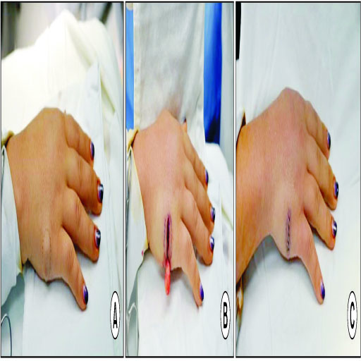
Fig. 1 (A) About 15 mm size ovoid shape well-defined firm soft tissue mass situated on subcutaneous fat, which is ulnar side of hand. (B) Under local anesthesia, excisional biopsy was performed. The incision line was designed along the long axis of mass for easy approach. Well encapsulated mass was excised. There is no involvement of neurovasular bundle. (C) Primary repair was performed using Nylon.
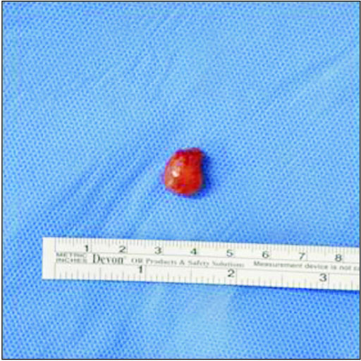
Fig. 2 Grossly, specimen was 15 mm size pinkish color oval shape mass with firm encapsulated characteristics.
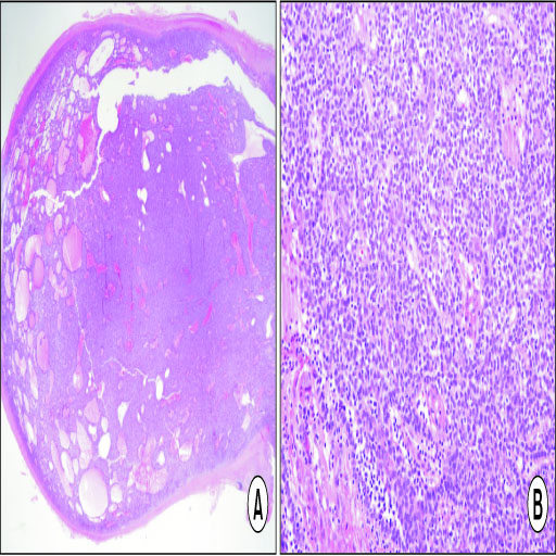
Fig. 3 (A) Well demarcated tumor with encapsulation is observed, not connected to epidermis (H&E stain, ×40). (B) In high power view, tumor is admixed with small basaloid, paler cells, and amount of lymphocytes (H&E stain, ×400). H&E: hematoxyling and eosin.
Cited by 1 articles
-
Painful eccrine spiradenoma containing nerve fibers in the forearm: a case report
Tae Hyun Kim, Seong Heum Jeong, Hyun Jeong Ha, Chung Hun Kim
Arch Hand Microsurg. 2024;29(1):60-63. doi: 10.12790/ahm.23.0053.
Reference
-
1. Padmini HR, Dinesh P. A rare and unique case of eccrine spiradenoma of the eyelid. J Evolution Med Dental Sci. 2013; 2:1700–1703.2. Al-Qattan MM. Eccrine spiradenoma of the hand clinically mimicking a lipoma. J Hand Surg. 1996; 21:551–552.
Article3. Alfonso-Trujillo I, Arteaga-Hernández E, Pérez-Suárez JC. Eccrine Spiradenoma in a zosteriform distribution: presentation of a case. Actas Dermosifiliogr. 2009; 100:619–620. Spanish.
Article4. Siegel HJ, Said-Al-Naief N, Long J, Lopez-Ben RR, Klein M. Giant eccrine spiradenoma of the hand. Am J Orthop. 2008; 37:E141–E143.5. Mambo NC. Eccrine spiradenoma: clinical and pathologic study of 49 tumours. J Cutan Pathol. 1983; 10:312–320.6. Noto G, Bongiorno MR, Pravià G, Aricò M. Multiple nevoid spiradenomas. Am J Dermatopathol. 1994; 16:280–284.
Article7. Gupta S, Radotra BD, Kaur I, Handa S, Kumar B. Multiple linear eccrine spiradenomas with eyelid involvement. J Eur Acad Dermatol Venereol. 2001; 15:163–166.
Article8. Amoroso C, Grandi E, Carinci F. Eccrine spiradenoma of the ear: case report. Int J Oral Maxillofac Surg. 2003; 32:662–663.
Article9. Criton S, Aravindan KP. Zosteriform network of spiradenoma. Indian J Dermatol Venereol Leprol. 1996; 62:185–186.10. Mirza I, Kloss R, Sieber SC. Malignant eccrine spiradenoma. Arch Pathol Lab Med. 2002; 126:591–594.
Article
