Ann Dermatol.
2018 Jun;30(3):392-393. 10.5021/ad.2018.30.3.392.
A Case of Pyoderma Gangrenosum with Myelodysplastic Syndrome
- Affiliations
-
- 1Department of Dermatology, Kangdong Sacred Heart Hospital, Hallym University College of Medicine, Seoul, Korea. hj2456hj@naver.com
- KMID: 2419195
- DOI: http://doi.org/10.5021/ad.2018.30.3.392
Abstract
- No abstract available.
Figure
-
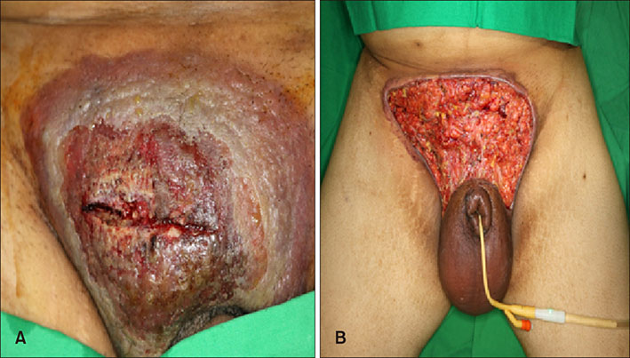
Fig. 1 (A) 15×5 cm sized, solitary large erythematous round plaque with raised border and surrounding violaceous patches on the suprapubia area. (B) 25×20 cm sized lesion, after necrotic wound debridement.
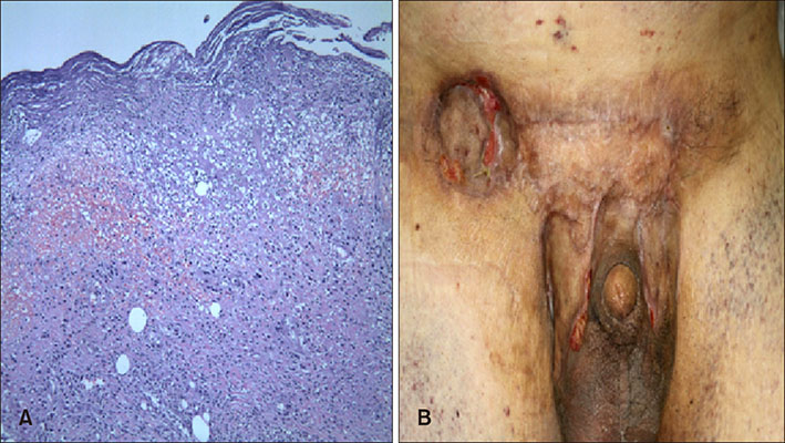
Fig. 2 (A) Skin biopsy specimen showed dermal and perivascular infiltration with several neutrophils and lymphohistiocytes (H&E, ×40). (B) Almost healed skin lesion following 5 month of cyclosporine and steroid therapy.
Reference
-
1. Lee JI, Park HJ, Lee JY, Cho BK. A case of pyoderma gangrenosum with ulcerative colitis treated with mesalazine. Ann Dermatol. 2010; 22:422–425.
Article2. von den. Pyoderma gangrenosum: a report of 44 cases with follow-up. Br J Dermatol. 1997; 137:1000–1005.
Article3. Yamanaka K, Kuniyuki S, Maekawa N, Yoshida Y, Teshima H. Pyoderma gangrenosum with myelodysplastic syndrome treated with analogous bone marrow transplantation. Acta Derm Venereol. 2009; 89:105–106.
Article4. Litvak D, Kirsner RS, Pakdaman NN, Federman DG. Pyoderma gangrenosum and myelodysplastic syndrome. South Med J. 2000; 93:923–925.
Article5. Koca E, Duman AE, Cetiner D, Buyukasik Y, Haznedaroglu IC, Uner A, et al. Successful treatment of myelodysplastic syndrome-induced pyoderma gangrenosum. Neth J Med. 2006; 64:422–424.
- Full Text Links
-
- Actions
-
Cited
- CITED
-
- Close
- Share
-
- Similar articles
-
- Nuclear segmentation anomaly of neutrophils in a case of pyoderma gangrenosum with myelodysplastic syndrome
- A Case of Successful Combination Therapy of Systemic Steroid and Topical 0.03% Tacrolimus for Pyoderma Gangrenosum in a Patient with Myelodysplastic Syndrome
- Unusual Cutaneous Neutrophilic Infiltration in Myelodysplasia Syndrome : A Nuclear Segmentation Anomaly
- A Case of Behcet's Disease Associated with Pyoderma Gangrenosum
- A Case of Pyoderma Gangrenosum Occurring in Behcet's Disease
