Simultaneous Closure of a Left Atrial Appendage through an Atrial Septal Defect and the Atrial Septal Defect
- Affiliations
-
- 1Division of Cardiology, Severance Cardiovascular Hospital, Yonsei University Health System, Seoul, Korea. kjs1218@yuhs.ac
- 2Cardiovascular Research Institute, Yonsei University College of Medicine, Seoul, Korea.
- KMID: 2418908
- DOI: http://doi.org/10.3349/ymj.2017.58.6.1237
Abstract
- Left atrial appendage (LAA) occlusion can be employed as an alternative treatment to oral anticoagulation in patients with atrial fibrillation to prevent embolic events. Atrial septal defect (ASD) may be related with right heart dysfunction and allow paradoxical embolism to occur. However, occlusion of both LAA through atrial access with ostium secundum ASD and ASD in the same setting is unusual. Therefore, we report a case in which a LAA and an ASD was sequentially occluded.
MeSH Terms
-
Aged
Atrial Appendage/diagnostic imaging/physiopathology/*surgery
Atrial Fibrillation/*complications/physiopathology
Heart Atria/physiopathology
Heart Septal Defects, Atrial/diagnostic imaging/physiopathology/*surgery
Humans
Male
Pyrazoles/*administration & dosage
Pyridones/*administration & dosage
*Septal Occluder Device
Thrombosis/*prevention & control
Tomography, X-Ray Computed
Treatment Outcome
Pyrazoles
Pyridones
Figure
-
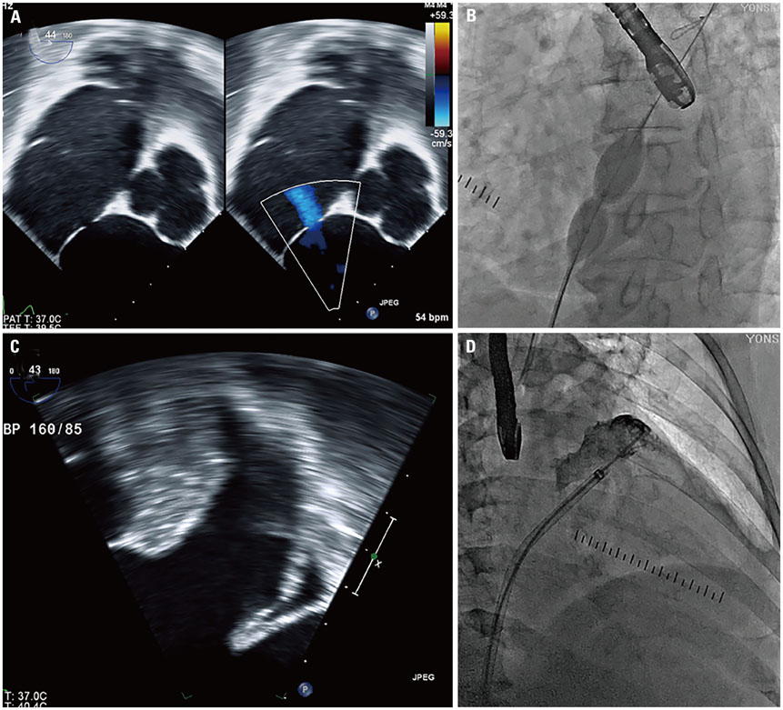
Fig. 1 Two dimensional TEE image of ASD (A). Fluoroscopic evaluation of ASD during expansion of the sizing balloon (B). TEE (C) and fluoroscopic images (D) of left atrial appendage before implantation. ASD, atrial septal defect; TEE, transesophageal echocardiographic.
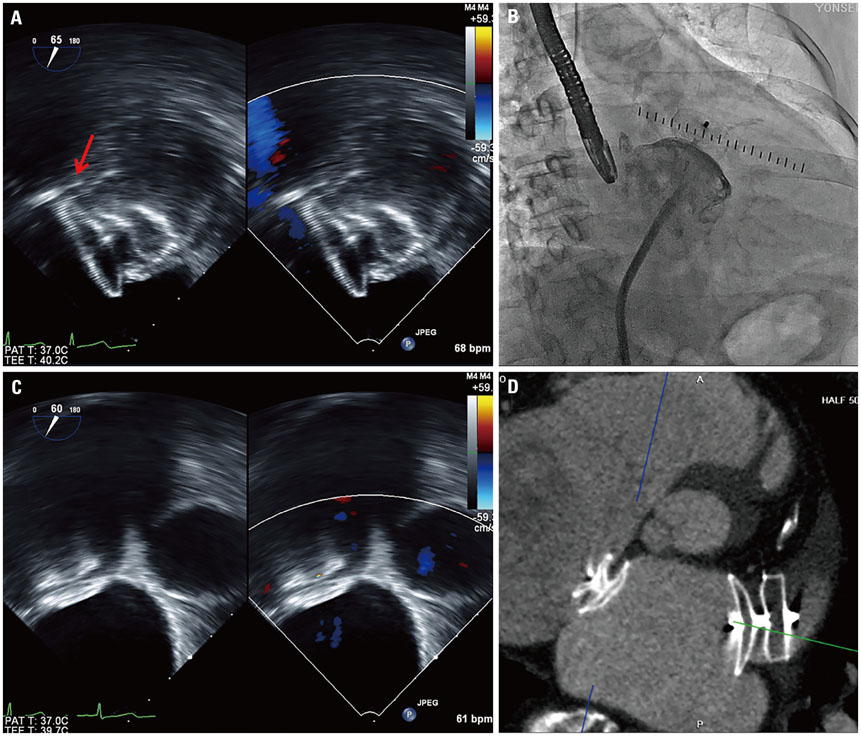
Fig. 2 The deployed left atrial appendage closure device (ACP 30 mm) was confirmed with transesophageal echocardiographic imaging (arrow: left circumflex artery) (A) and fluoroscopic imaging (B). The Amplatzer septal occluder (11 mm) was located in the proper position, which was confirmed by transesophageal echocardiography (C). After the procedure, implanted Amplatzer septal occluder (11 mm) and ACP (30 mm) demonstrated by heart computed tomography (D). ACP, Amplatzer Cardiac Plug.
Reference
-
1. Kim YL, Joung B, On YK, Shim CY, Lee MH, Kim YH, et al. Early experience using a left atrial appendage occlusion device in patients with atrial fibrillation. Yonsei Med J. 2012; 53:83–90.
Article2. Meier B, Blaauw Y, Khattab AA, Lewalter T, Sievert H, Tondo C, et al. EHRA/EAPCI expert consensus statement on catheter-based left atrial appendage occlusion. Europace. 2014; 16:1397–1416.
Article3. Koermendy D, Nietlispach F, Shakir S, Gloekler S, Wenaweser P, Windecker S, et al. Amplatzer left atrial appendage occlusion through a patent foramen ovale. Catheter Cardiovasc Interv. 2014; 84:1190–1196.
Article4. Tzeis S, Andrikopoulos G, Deisenhofer I, Ho SY, Theodorakis G. Transseptal catheterization: considerations and caveats. Pacing Clin Electrophysiol. 2010; 33:231–242.
Article5. Alkhouli M, Sarraf M, Zack CJ, Holmes DR, Rihal CS. Iatrogenic atrial septal defect following transseptal cardiac interventions. Int J Cardiol. 2016; 209:142–148.
Article6. Singh SM, Douglas PS, Reddy VY. The incidence and long-term clinical outcome of iatrogenic atrial septal defects secondary to transseptal catheterization with a 12F transseptal sheath. Circ Arrhythm Electrophysiol. 2011; 4:166–171.
Article7. Berthet K, Lavergne T, Cohen A, Guize L, Bousser MG, Le Heuzey JY, et al. Significant association of atrial vulnerability with atrial septal abnormalities in young patients with ischemic stroke of unknown cause. Stroke. 2000; 31:398–403.
Article8. Handke M, Harloff A, Olschewski M, Hetzel A, Geibel A. Patent foramen ovale and cryptogenic stroke in older patients. N Engl J Med. 2007; 357:2262–2268.
Article9. Lechat P, Mas JL, Lascault G, Loron P, Theard M, Klimczac M, et al. Prevalence of patent foramen ovale in patients with stroke. N Engl J Med. 1988; 318:1148–1152.
Article10. Warnes CA, Williams RG, Bashore TM, Child JS, Connolly HM, Dearani JA, et al. ACC/AHA 2008 guidelines for the management of adults with congenital heart disease: A Report of the American College of Cardiology/American Heart Association Task Force on Practice Guidelines (Writing Committee to Develop Guidelines on the Management of Adults With Congenital Heart Disease). Developed in Collaboration with the American Society of Echocardiography, Heart Rhythm Society, International Society for Adult Congenital Heart Disease, Society for Cardiovascular Angiography and Interventions, and Society of Thoracic Surgeons. J Am Coll Cardiol. 2008; 52:e143–e263.11. Gafoor S, Franke J, Boehm P, Lam S, Bertog S, Vaskelyte L, et al. Leaving no hole unclosed: left atrial appendage occlusion in patients having closure of patent foramen ovale or atrial septal defect. J Interv Cardiol. 2014; 27:414–422.
Article
- Full Text Links
-
- Actions
-
Cited
- CITED
-
- Close
- Share
-
- Similar articles
-
- A Case of Atrial Septal Defect in Identical Twins
- A Case of Atrial Septal Aneurysm Associated with Atrial Septal Defect
- Subacute, Silent Embolization of Amplatzer Atrial Septal Defect Closure Device to the Pulmonary Artery
- Transcatheter Closure of Secundum Atrial Septal Defect with the Amplatzer Septal Occluder
- Procedural, Early and Long-Term Outcomes after Transcatheter Atrial Septal Defects Closure: Comparison between Large and Very Large Atrial Septal Defect Groups
